Navigating the Frailty Factor in Atrial Fibrillation Management

In a new editorial, researchers discuss the impact of frailty on clinical decision-making in managing patients with atrial fibrillation and the prescription of oral anticoagulants.

Atrial fibrillation (AF) is a type of heart arrhythmia that occurs when the ​​heart’s electrical signals become irregular. This condition can increase the risk of stroke and heart failure, and becomes more common in older adults. Frailty is another condition that coincides with aging. Frailty encompasses an accumulation of deficits and can be defined as decreased physical function and resilience. For a significant proportion of the elderly population, both of these conditions coexist. This convergence can lead to additional health issues and further complicate the clinical landscape for aging individuals. 

“The impact of frailty on outcomes has not been previously well characterized in populations with AF.”

Studies on frailty and its overall impact on patients with AF are needed. In a new editorial paper, researchers Stephanie L. Harrison, Søren P. Johnsen and Gregory Y.H. Lip from Liverpool John Moores University and Liverpool Heart and Chest Hospital discuss some of the existing studies assessing frailty and AF in terms of patient outcomes and clinical decision-making. The researchers also discuss recommendations for clinical management of AF and frailty patients and include suggestions for future studies. On July 19, 2023, their editorial was published in Aging’s Volume 15, Issue 14, entitled, “The impact of frailty on the management of atrial fibrillation.”

Oral Anticoagulants for AF With Frailty: Yay or Nay?

Oral anticoagulants (OACs) are often prescribed to AF patients, as OACs aid in reducing blood clots and the risk of stroke. However, frailty patients are more susceptible to falls and therefore, more susceptible to bleeding risk. The prevention of blood clots is less than ideal for wound healing and can lead to impaired tissue repair, increased risk of infection and prolonged recovery times. Has frailty status impacted clinical decision-making when it comes to the prescription of OACs for AF patients?

“The impact of frailty on clinical decision making for managing patients with AF such as the prescription of oral anticoagulants (OACs) is unclear.”

In an effort to begin answering this question, the authors of this editorial reviewed a number of studies on frailty and AF. Some cross-sectional analyses revealed a significant association between frailty status and the prescription of OACs, while other studies found no such association. The authors explain that these dueling results may be due to differences in study populations, the tools/methods used to assess frailty and the timing of when the studies were conducted. The prescription of anticoagulants increased when a new class of OACs was introduced to the market.

Non-vitamin K antagonist oral anticoagulants (NOACs) were first introduced in the early 2010s. They are a class of drugs with significant advantages over traditional OACs, including less off-target effects, more predictable pharmacokinetics and a more targeted mechanism of action — making them a safer option for use in combination with other drugs. Notably, certain NOACs have specific reversal agents available, which can be used to rapidly reverse their anticoagulant effect in case of emergency. International guidelines recommended their use as a first-line treatment to reduce the risk of stroke in AF patients.

“Frailty alone should not be reason to withhold oral anticoagulation in patients with AF. In patients with frailty, the benefits of oral anticoagulation outweigh the small absolute risk of bleeding [6].”

Conclusions & Future Directions

The authors close the editorial by stating that additional research is needed to understand how frailty impacts clinical decision-making for the management of atrial fibrillation. They strongly suggest that frailty not be the sole reason AF patients are not prescribed potentially life-saving oral anticoagulants. There are more sensitive options available today, such as NOACs, that can potentially mitigate the drawbacks of anticoagulants for people with frailty. The authors also emphasize that pre-frailty assessments should be conducted for AF patients in order to reduce treatment risk and optimize the management of this condition.

“Assessment of pre-frailty in patients with AF and the development of strategies to address potentially modifiable components of pre-frailty to reduce the risk of frailty progression could have important implications to optimize care.”

Click here to read the full editorial published in Aging.

Aging is an open-access, peer-reviewed journal that has been publishing high-impact papers in all fields of aging research since 2009. These papers are available to readers (at no cost and free of subscription barriers) in bi-monthly issues at Aging-US.com.

Click here to subscribe to Aging publication updates.

For media inquiries, please contact [email protected].

Exploring the Impact of Cognitive Reserve on Cognitive Resilience

In a new editorial, researchers discuss their recent study investigating the effect that cognitive reserve has on brain integrity and cognitive resilience.

Why do some individuals maintain cognitive abilities throughout their lifespan and others do not? The better question may be: How can certain individuals preserve their cognitive abilities and delay the onset of dementia despite the presence of significant neuropathologies that would otherwise suggest cognitive decline? This question remains unanswered.

“What contributes to this ‘resilience’ [3], that is why some successfully cope with progressive neuropathology while others cannot tolerate the same level of neurodegeneration, is not fully understood.”

This unanswered question has driven researchers to consider the idea of “cognitive reserve.” The rather new concept of cognitive reserve suggests that certain factors, such as engaging in education, mental stimulation and challenging activities, can create a buffer against cognitive decline and delay the onset of cognitive impairment or dementia. Researchers continue to study cognitive reserve to better understand its mechanisms and potential implications for maintaining brain health and designing effective interventions.

In a new editorial paper, researchers Monica E. Nelson, Ross Andel and Jakub Hort from the University of South Florida’s​​ School of Aging Studies discussed the outcomes, lessons and future implications of their previous 2022 study. The team examined the influence of cognitive reserve proxies on the relationship between brain integrity and cognition. On July 14, 2023, their editorial was published in Aging’s Volume 15, Issue 13, entitled, “Cognitive reserve, neuropathology, and progression towards Alzheimer’s disease.”

Cognitive Reserve’s Effect on Brain Integrity and Cognitive Performance

In their 2022 study, a total 570 older adult participants were assessed from the Czech Brain Aging Study (a longitudinal cohort study from two memory clinics in the Czech Republic). Most of the participants (n = 457) were without dementia (including those with subjective cognitive decline and amnestic mild cognitive impairment) and the remaining participants were with dementia syndrome (n = 113). The researchers examined the influence of education and occupational position (cognitive reserve proxies) on the relationship between the participants’ hippocampal or total gray matter volume and cognitive performance. Measurements included brain volume, executive control, language, memory, attention/working memory, and visuospatial skills.

“[…] we assessed the inter-link between cognitive reserve, neuropathology, and cognitive functioning among participants with subjective cognitive decline, mild cognitive impairment, and dementia.”

The team found that the association between brain volume and cognitive performance varies based on cognitive reserve. Findings showed that a higher education and occupational position magnified the associations between brain volume and cognitive performance in participants without dementia. In participants with dementia, higher education decreased the associations between brain volume and visuospatial skills. Overall, the results showed that cognitive reserve affects the relationship between brain volume and cognitive performance, with greater cognitive reserve related to a stronger link before dementia diagnosis and a weaker link after.

Future Directions 

In their subsequent editorial, the researchers were forthcoming about limitations of this study and addressed key opportunities for future studies. Limitations were identified as the use of a relatively homogeneous sample population, the absence of the use of biomarkers in diagnosis and the cross sectional design. Cross-sectional studies may not fully capture disease-related changes in neuropathology and could present a distorted view of the linkages between cognitive reserve, neuropathology and cognitive outcomes. The authors advocate for conducting longitudinal studies to track how cognitive reserve operates in individuals as they progress from normal to dementia. 

Additionally, the team wrote that future studies would be improved by investigating a range of Alzheimer’s disease biomarkers, such as beta-amyloid and tau, individually and together, to understand how they influence the associations between cognitive reserve, brain health and cognition. Different biomarkers may lead to varied results in how cognitive reserve moderates these associations. And finally, future studies should also assess older adults across the cognitive spectrum to determine when cognitive reserve is protective against brain health decline and neuropathology, and when its effectiveness diminishes. Some researchers have suggested a U-shaped relationship to explain mixed findings in different studies.

“Even though our study represents one of the first to come from Eastern Europe [4], future work should be conducted in additional populations, representing geographic, racial, and socioeconomic diversity.”

Implications

The potential impact of this research may be important, as it could lead to the development of effective interventions and strategies to preserve cognitive abilities and delay the onset of dementia. By gaining a deeper understanding of cognitive reserve and its mechanisms, we can take steps to promote brain health throughout life, potentially reducing the burden of dementia on individuals, communities and society overall.

As research continues in this field, it is clear that cognitive reserve holds great promise for unlocking the secrets of cognitive resilience and paving the way for healthier aging and improved quality of life for older adults. By addressing the limitations of current studies and exploring new avenues of investigation, we move closer to finding answers to the vital question of how some individuals maintain their cognitive abilities despite the presence of significant neuropathologies, while others do not.

“By assessing cognitive reserve in distinct populations, a more complete understanding of how cognitive reserve relates to neuropathology and cognition and whether these associations may be affected by distinct macro-level contextual differences among populations can be established. Disentangling these complex relationships may provide a critical step in reducing the impact of dementia on society.”

Click here to read the full editorial published by Aging.

Aging is an open-access, peer-reviewed journal that has been publishing high-impact papers in all fields of aging research since 2009. These papers are available to readers (at no cost and free of subscription barriers) in bi-monthly issues at Aging-US.com.

Click here to subscribe to Aging publication updates.

For media inquiries, please contact [email protected].

Can a Leaky Gut Lead to Alzheimer’s Disease?

In a new editorial, researchers explore how a leaky gut can lead to Alzheimer’s disease using the Seed and Soil Model of Neurocognitive Disorders to explain.

New research continues to illuminate the far-reaching implications of the gut microbiome and its crucial role in our overall health. The term “gut dysbiosis” refers to an imbalance of healthy and unhealthy microbes in the gastrointestinal tract. Repercussions of gut dysbiosis are not only limited to innocuous discomfort—it can lead to immune dysregulation and trigger a cascade of various disease states. 

In a new editorial paper, researchers Chun-Che Hung, Kristi M. Crowe-White and Ian M. McDonough from Chang Gung University and The University of Alabama discuss the relationship between gut dysbiosis and neurocognitive disorders such as Alzheimer’s disease (AD). Their editorial was published in Aging’s Volume 15, Issue 12, on June 19, 2023, entitled, “A seed and soil model of gut dysbiosis in Alzheimer’s disease.”

“[…] recent research has demonstrated a crucial role of gut microbiota in the etiopathogenesis of AD [Alzheimer’s disease] that offers a new window into possible origins and consequences of AD through interactions between gut microbiota and the central nervous system, known as the ‘microbiota-gutbrain axis’ [1].”

The Seed and Soil Model of Neurocognitive Disorders

The “Seed and Soil Model” in biology was first used in an attempt to describe why some individuals who are predisposed to developing neurocognitive disorders do not ever develop them. As the researchers wrote in their editorial, the “seeds” in this analogy represent genetic predispositions or a family history of a particular disease state. The “soil” represents the external environment that either enables or disables the expression of these seeds. This external environment can be influenced by behavioral and/or lifestyle factors. Although this model did not originally include the microbiota-gut-brain axis, the authors of this editorial are now applying it.

Interestingly, the researchers here have related the “leaky gut” phenomenon of gut dysbiosis to Alzheimer’s disease (AD). A leaky gut, plainly described as increased intestinal permeability, is a condition where the lining of the intestine becomes more porous. This allows larger molecules and toxins to pass through into the bloodstream—opening the door to potential inflammation and various health problems. 

Metabolites involved with gut leakiness have previously been linked to increased permeability of the blood-brain barrier (BBB). The opening of the BBB allows bacterial endotoxins to travel from the gut to the brain environment. This can increase inflammation within the system. The authors propose that gut leakiness, through the Seed and Soil Model, may explain why some people predisposed to AD realize the disease, while those without gut dysbiosis may not.

“According to the Seed and Soil Model of Neurocognitive Disorders, this translocation would create a toxic microenvironment in the brain vulnerable to pathogenesis, especially for those with a genetic predisposition to AD.”

Conclusion

“According to the Seed and Soil Model of Neurocognitive Disorders, environmental and behavioral patterns can influence the balance of neuroprotection vs. toxicity of the brain’s micro-environment.”

In sum, emerging research continues to shed light on the significance of the gut microbiome and its connection to our overall health. The editorial by Hung, Crowe-White and McDonough explores the relationship between gut dysbiosis and neurocognitive disorders, particularly Alzheimer’s disease, through the lens of the Seed and Soil Model of Neurocognitive Disorders. By considering the impact of leaky gut and the translocation of bacterial endotoxins on the brain, the authors propose that gut dysbiosis may contribute to the pathogenesis of AD, particularly in individuals with a genetic predisposition. This perspective opens new avenues for understanding the complex interactions within the microbiota-gut-brain axis and provides insights into ways to potentially stave off cognitive decline with diet and lifestyle interventions.

“Here, we extend the model to better understand how the microbiota-gut-brain axis may play a causal role in the development of AD. However, more research is needed to test additional hypotheses of the model.”

Click here to read the full research paper published by Aging.

Aging is an open-access, peer-reviewed journal that has been publishing high-impact papers in all fields of aging research since 2009. These papers are available to readers (at no cost and free of subscription barriers) in bi-monthly issues at Aging-US.com.

Click here to subscribe to Aging publication updates.

For media inquiries, please contact [email protected].

How Telomere Dysfunction Affects Female Fertility (A Mouse Study)

In a new study, researchers aimed to reveal a link between telomere dysfunction, ovarian aging and infertility using a mouse model of accelerated-reproductive senescence.

Telomeres are the protective caps at the ends of chromosomes that prevent DNA damage and maintain genomic stability. However, telomeres shorten with each cell division and eventually reach a critical length that triggers cellular senescence or death. Telomere length (TL) and telomerase activity (TA), the enzyme that replenishes telomeric repeats, are influenced by genetic and environmental factors and vary among tissues and individuals.

“Telomere attrition has been identified as one of the molecular determinants of aging [7].”

Telomere dysfunction has been implicated in various age-related diseases, including infertility. Ovarian aging is the main cause of infertility in women, as it leads to a decline in both the quantity and quality of oocytes. Previous studies have shown that TL and TA are reduced in oocytes and granulosa cells of women with diminished ovarian reserve or poor response to ovarian stimulation. Moreover, TL and TA have been associated with ovarian reserve markers and pregnancy outcomes in assisted reproductive technologies.

To better understand the molecular mechanisms underlying ovarian aging and infertility, researchers Alba M. Polonio, Marta Medrano, Lucía Chico-Sordo, Isabel Córdova-Oriz, Mauro Cozzolino, José Montans, Sonia Herraiz, Emre Seli, Antonio Pellicer, Juan A. García-Velasco, and Elisa Varela from The Health Research Institute La Fe (IIS La Fe), IVIRMA Rome, New Jersey and Madrid, Centro Anatomopatológico, Yale School of Medicine, University of Valencia, and Rey Juan Carlos University conducted a new study using a mouse model of accelerated aging: the Senescence-Accelerated Mouse Prone 8 (SAMP8). On May 23, 2023, their research paper was published in Aging’s Volume 15, Issue 11, entitled, “Impaired telomere pathway and fertility in Senescence-Accelerated Mice Prone 8 females with reproductive senescence.”

The Study

The SAMP8 mouse model, which has previously been suggested as an Alzheimer’s disease model of aging, also exhibits a shortened estrous cycle, elevated follicle-stimulating hormone (FSH) levels, and reduced fertility in females at just seven months of age. SAMP8 mice have a shorter lifespan compared to senescence-accelerated mouse resistant 1 (SAMR1) mice. SAMR1 mice do not exhibit reproductive senescence. Thus, the researchers deemed the SAMR1 mouse model an appropriate control group to study the SAMP8 mouse model as a model of ovarian aging and infertility. 

“In the current study, we sought to investigate whether the SAMP8 mice, which show accelerated-reproductive senescence, have alterations in their telomere pathway. This question has not yet been explored in relation to reproduction in this model.”

In this study, the team compared the TL and TA in blood and ovary samples from the SAMP8 female mice at seven months of age (when they show signs of reproductive senescence) with age-matched control SAMR1 mice. They also evaluated the ovarian follicle development, the expression of telomerase subunits (TERT and TERC), and the reproductive outcomes after ovarian stimulation in both groups of mice. In sum, the researchers measured survival rates (in male and female mice), alteration in the telomere pathway at seven months of age, TERT and TERC expression levels, TA on the TL of granulosa cells in developing follicles, and impairment/alterations in the telomere pathway in oogenesis and embryo development.

The results revealed that SAMP8 females had a reduced median lifespan compared to SAMP8 males and SAMR1 males and females. In blood, SAMP8 females had lower mean TL and higher accumulation of short telomeres than the other mice. In ovary, SAMP8 females had lower TA and TERT expression. Furthermore, SAMP8 females had fewer primordial, primary, secondary, and antral follicles than control females, indicating a diminished ovarian reserve. After ovarian stimulation, SAMP8 females had a lower number of oocytes than controls of the same age. Their results suggested that oogenesis and embryo development is impaired in SAMP8 mice at seven months compared to age-matched controls, and this coincides with alterations in the telomere pathway.

Conclusions

“Thus, SAMP8 females represent a bona fide model for the analysis of fertility, not only because it shows similar phenotype to middle-aged women as stated earlier [43], but also because the alterations in the telomere pathway are found in women with fertility disorders [37, 38, 40, 41] and this pathway links reproduction with longevity.”

The researchers concluded that SAMP8 females have impaired telomere pathway and fertility, reflecting signs of reproductive senescence described in middle-aged women. They suggested that the SAMP8 model could be useful in studying the role of telomere dysfunction in ovarian aging and infertility. In addition, this mouse model could be used to test potential therapeutic interventions to improve female reproductive health.

“Understanding the molecular pathways underlying aging and fertility, provides a basis for further studies focused on several topics. First, the analysis of embryo alterations, which can be better assessed in mice than in humans. Second, how reproductive lifespan improvement may ameliorate elderly health. And third, the mechanisms underlying follicle recruitment and development, which are not completely known.”

Click here to read the full research paper published by Aging.

Aging is an open-access, peer-reviewed journal that has been publishing high-impact papers in all fields of aging research since 2009. These papers are available to readers (at no cost and free of subscription barriers) in bi-monthly issues at Aging-US.com.

Click here to subscribe to Aging publication updates.

For media inquiries, please contact [email protected].

The Impact of Age, Sex, CMV, and Smoking on Circulating Immune Cells

In a new study, researchers investigated associations between circulating immune cells and age, sex, CMV infection, and smoking.

The Impact of Age, Sex, CMV, and Smoking on Circulating Immune Cells

As we age, our immune system undergoes changes that influence our susceptibility to various diseases. Certain factors, such as smoking, viruses, age, and sex can have differential impacts on our various circulating immune cells. How changes to these immune cells contribute to cardiovascular disease and other age-related diseases is not yet fully understood. More research is needed to fully understand the underlying mechanisms and implications. 

“Understanding the composition of circulating immune cells with aging and the underlying biologic mechanisms driving aging may provide molecular targets to slow the aging process and reduce age-related disease.”

In a new study, researchers Yuan Fang, Margaret F. Doyle, Jiachen Chen, Jesse Mez, Claudia L. Satizabal, Michael L. Alosco, Wei Qiao Qiu, Kathryn L. Lunetta, and Joanne M. Murabito from Boston University, Boston Medical Center, University of Vermont, and University of Texas Health Science Center aimed to characterize the circulating innate and adaptive immune system by profiling immune cell phenotypes from a community-based cohort. Their research paper was published in Aging’s Volume 15, Issue 10, on April 27, 2023, entitled, “Circulating immune cell phenotypes are associated with age, sex, CMV, and smoking status in the Framingham Heart Study offspring participants.”

“We hypothesize that we will identify immune cell phenotype and ARIP [age-related immune phenotype] measure associations with CMV serostatus, age, and sex, as well as associations with cardiovascular risk factors.”

The Study and Participant Characteristics

The Framingham Heart Study (FHS) is a community-based prospective cohort study that began in 1948. It initially recruited 5,209 primarily white American adults of European ancestry as the Original cohort. In 1971, the Offspring cohort was established, consisting of the children of the Original cohort and their spouses. The Offspring participants have been examined every 4-8 years since enrollment. 

For this study, 1,332 Offspring participants who attended exam seven (1998 to 2001) and had two or more vials of stored peripheral blood mononuclear cells (PBMCs) were identified. From this group, a study sample of 996 dementia-free individuals, aged 40 years and older, was selected. This cohort had a mean age of 62 years, with 52% representing males. All participants provided written informed consent, and the FHS exams were approved by the Institutional Review Board at Boston University Medical Center.

The research team used cryopreserved cell samples from the study participants to conduct comprehensive analyses of 116 circulating immune cell phenotypes, including subtypes of CD4 and CD8 T cells, B cells, NK cells, and monocytes. These subsets were further categorized based on specific surface markers to provide a detailed characterization of the immune cell populations.

The Results

Significant associations between circulating immune cell phenotypes and age, sex, a common virus, and smoking were revealed in this study. With advancing age, researchers saw a decline in the overall number of immune cells, as well as alterations in the distribution of different immune cell subsets. Notably, older individuals exhibited a higher proportion of memory T cells and a lower proportion of naive T cells, suggesting a shift towards a more experienced immune profile. Furthermore, females exhibited a higher abundance of immune cells compared to males, which may contribute to their generally stronger immune responses.

Cytomegalovirus (CMV), a common herpesvirus, can have a profound impact on the immune system. The study found that CMV seropositivity was associated with distinct alterations in immune cell phenotypes. CMV-positive individuals displayed higher numbers of late-stage differentiated effector memory T cells, which are indicative of previous exposure to CMV. This observation suggests that CMV infection contributes to the age-related changes in immune cell populations.

Smoking has long been recognized as a detrimental habit that affects overall health, including the immune system. This study uncovered compelling evidence linking smoking status to immune cell phenotypes. Smokers exhibited a higher proportion of pro-inflammatory immune cells, such as activated T cells and pro-inflammatory monocytes, while non-smokers had a higher proportion of regulatory T cells that help maintain immune balance. These findings emphasize the detrimental impact of smoking on immune cell profiles and further underscore the importance of smoking cessation.

“Importantly, we did not identify significant immune cell associations with other risk factors, such as body mass index, prevalent cardiovascular disease, hypertension or diabetes.”

Conclusions

“Our observations confirm and extend known associations of immune cell subtypes with CMV and age that show a shift from a naïve phenotype towards an exhausted phenotype. We report sex differences, with males exhibiting a more exhausted, cytotoxic landscape than females. We identified associations between CD8 exhausted cells and B cell subsets, but not overall B cells, with smoking status.” 

This research provides valuable insights into the relationship between circulating immune cell phenotypes and age, sex, CMV infection, and smoking status. In conclusion, the team did not find significant associations between these immune cells and cardiovascular risk factors. They did find some weak associations with cardiovascular disease, diabetes and hypertension. The findings contribute to our understanding of age-related changes in the immune system and highlight the impact of lifestyle factors on immune health. By unraveling the complex interplay between these variables, this study paves the way for future research on interventions and strategies to support healthy immune aging.

“While further studies in larger, more diverse sample[s] and more than one time point with immunophenotypic data are needed, this work will provide a valuable resource for future studies of the association of immune cell phenotypes and incident age-related disease.”

Click here to read the full research paper published by Aging.

Aging is an open-access, peer-reviewed journal that has been publishing high-impact papers in all fields of aging research since 2009. These papers are available to readers (at no cost and free of subscription barriers) in bi-monthly issues at Aging-US.com.

Click here to subscribe to Aging publication updates.

For media inquiries, please contact [email protected].

Brain Aging Insights from Individuals Without Neurodegeneration

The Trending With Impact series highlights Aging publications (listed by MEDLINE/PubMed as “Aging (Albany NY)” and “Aging-US” by Web of Science) that attract higher visibility among readers around the world online, in the news and on social media—beyond normal readership levels. Look for future science news about the latest trending publications here, and at Aging-US.com.

Listen to an audio version of this article

A healthy brain continuously produces new proteins to support synaptic plasticity, maintain neuronal health, facilitate signaling pathways, produce neurotransmitters, enable neuroplasticity and adaptation, and meet its metabolic demands. These processes are essential for normal brain function, learning, memory, and overall cognitive abilities. Researchers believe that the dysregulation of proteins is at the core of brain aging. However, the exact recipe for protein dysregulation that leads to accelerated brain aging and neurodegenerative disorders has yet to be brought to light. 

Previous brain proteostasis (referring to the maintenance of protein homeostasis in brain cells) studies in individuals with Alzheimer’s disease (AD) pathology and age-related neuropathological changes have shown protein dysregulation leading to a buildup of amyloid plaques and neurofibrillary tangles. While these studies have greatly enhanced our knowledge of brain aging, gaps in our understanding remain. What proteomic characteristics do healthy brain aging individuals—without neurodegenerative disorders—have in common?

“To our knowledge, whole phosphoproteomes centered on the human brain aging without AD pathology are unavailable.”

In a new study, researchers Pol Andrés-Benito, Ignacio Íñigo-Marco, Marta Brullas, Margarita Carmona, José Antonio del Rio, Joaquín Fernández-Irigoyen, Enrique Santamaría, Mónica Povedano, and Isidro Ferrer from Bellvitge Institute for Biomedical Research, Universidad Pública de Navarra, Barcelona Institute for Science and Technology, and University of Barcelona aimed to shed light on the mechanisms underlying brain aging in the absence of AD pathology and age-related neuropathological changes. Their research paper was published on May 13, 2023, in Aging’s Volume 15, Issue 9, and entitled, “Proteostatic modulation in brain aging without associated Alzheimer’s disease-and age-related neuropathological changes.”

The Study

The production of new proteins is crucial for maintaining protein homeostasis in the brain. A post-translational modification used to maintain this homeostasis is protein phosphorylation. In this study, the researchers conducted proteomic and phosphoproteomic analyses of frontal cortex samples from the donor brains of deceased individuals between the ages of 30 and 85. These individuals had passed away due to non-neurological complications and were reported to have had full cognitive function. Individuals were divided into four groups: young group one (30–44), middle-aged group two (45-52), early-elderly group three (64–70), and late-elderly group four (75–85).

“​​We chose the FC [frontal cortex] because of its role in cognition and emotion and the abundant molecular information that permits comparison with other studies.”

Conventional label-free- and SWATH- (sequential window acquisition of all theoretical fragment ion spectra) mass spectrometry were used to assess the (phospho)proteomes of the frontal cortices from individuals in all four age groups. Immunohistochemistry and/or western blotting was/were also used to validate a subgroup of proteins. The researchers categorized deregulated proteins and phosphoproteins into eight clusters based on their age-dependent expression similarity (see paper for clusters). Interestingly, protein and phosphoprotein levels of the larger hierarchical clusters were stable until the age of 70 years. After 70, the late-elderly group showed significant decreased or increased expression of protein clusters one and seven, and major phosphorylation modifications occurred in clusters four and eight.

Results

The team then used multi-comparative analyses to categorize altered proteins and phosphoproteins as neuronal, astroglial, oligodendroglial, microglial, and endothelial. They observed a similar pattern among proteomic and phosphoproteomic alterations: major changes were related to neuronal cell populations across all four groups—and these changes were more pronounced with age. Cytoskeletal and membrane proteins accounted for the largest number of differentially-expressed proteins and phosphoproteins.

“Furthermore, main alterations in the proteome are associated with proteins specific to neuronal populations, rather than those found in other cell types in the brain.”

Their findings also revealed a decline in the expression of P20S α + β with aging, while the expression of P19S and immunoproteasome subunits LMP2 and LMP7 remained preserved. Notably, the expression levels of an autophagy component, ATG5, remained unchanged with age. Some mitochondrial membrane proteins showed altered levels at advanced ages, but key markers of mitochondrial function were preserved. These findings suggest a potential preservation of these pathways in advanced aging, contrasting with observations in neurodegenerative disorders. Additionally, reduced levels of GSK3α/β were observed, and the researchers point out that this decrease in GSK3α/β with age may be understood as protective against different age-related brain diseases.

Summary & Conclusion

“Therefore, our results fill the gap between brain ageing without ADNC [AD neuropathological changes], and cases with early and advanced stages of AD pathology.”

The researchers are forthcoming about limitations of this study. Given it is rare for old-aged individuals not to have neurological deficits, AD or other neuropathological changes, their main limitation was that each of the four groups included merely four individuals. Despite limitations, these findings contribute to our understanding of brain aging in the absence of AD pathology and age-related neuropathological changes. 

The study revealed that major changes in protein expression were primarily associated with neuronal cell populations and became more pronounced with age. The preservation of specific protein pathways, proteasome components, autophagy-related components, and mitochondrial markers in advanced aging individuals without neurodegenerative disorders suggests the presence of resilience mechanisms that protect against protein dysregulation and neurodegeneration. Overall, this research provides valuable insights into the proteomic characteristics of healthy brain aging and highlights potential targets for therapeutic interventions aimed at promoting healthy brain aging and preventing age-related neurodegenerative diseases. Further studies are necessary to elucidate the specific mechanisms underlying these proteomic alterations and their functional implications in brain aging.

“The present observations identify proteostatic changes, including different changes in the phosphoproteome in the human FC in brain aging in the rare subpopulation of old-aged individuals without neurological deficits, and not having ADNC and other neuropathological change in any region of the telencephalon.”

Click here to read the full research paper published by Aging.

Aging is an open-access, peer-reviewed journal that has been publishing high-impact papers in all fields of aging research since 2009. These papers are available to readers (at no cost and free of subscription barriers) in bi-monthly issues at Aging-US.com.

Click here to subscribe to Aging publication updates.

For media inquiries, please contact [email protected].

The Brain Age Gap

The Trending With Impact series highlights Aging publications (listed by MEDLINE/PubMed as “Aging (Albany NY)” and “Aging-US” by Web of Science) that attract higher visibility among readers around the world online, in the news and on social media—beyond normal readership levels. Look for future science news about the latest trending publications here, and at Aging-US.com.

Listen to an audio version of this article

Aging is a risk factor for many diseases, including Alzheimer’s disease (AD). While scientists have made some progress in understanding the physiology of aging and its relationship to AD and related disorders, our understanding remains incomplete (to say the least). It is possible that civilization is currently in the midst of an artificial intelligence (AI) and machine learning (ML) “boom.” Researchers are now using AI and ML technologies to elevate our comprehension of aging and aging-related diseases.

“Artificial intelligence (AI) and machine learning (ML) technologies can help us better understand these diseases and aging itself by using biological data from the brain or other sources to create a mapping between age and biological data.”

In a new editorial paper, researchers Jeyeon Lee, Leland R. Barnard and David T. Jones from the Mayo Clinic in Rochester, Minnesota, discuss a recent study they conducted and explore the potential of AI to revolutionize the field of geriatrics. Their editorial was published in Aging’s Volume 15, Issue 8, on April 3, 2023, entitled, “Artificial intelligence and the aging mind.”

Their Study

In a recent 2022 study, Lee, Barnard, Jones, and the rest of their team developed convolutional neural network-based brain age prediction models using a large collection of data from brain magnetic resonance imaging (MRI) and brain fluorodeoxyglucose positron-emission tomography (FDG-PET) in people aged from 26 to 98 years old. In a sample of cognitively normal individuals, the AI models showed accurate brain age estimation of which a mean absolute error (MAE; unit, years) was 3.08±0.14 for the FDG-based model and 3.49±0.16 for the MRI-based model. 

The team found that higher brain age gaps (the difference between biological age and chronological age) were estimated in cohorts with neurodegenerative disorders—including mild cognitive impairment (MCI), AD, frontotemporal dementia (FTD), and dementia with Lewy bodies (DLB)—than normal controls. The brain age gap was strongly associated with pathologic tau protein levels and cognitive test scores. This gap also showed longitudinal predictive ability for cognitive decline in AD-related disorders.

“Interestingly, the brain imaging patterns generating brain age gaps in AD showed higher similarity with normal aging than other neurodegenerative syndromes implying that AD might be more like an accelerated representation of biological aging than others.”

Summary & Conclusions

The study conducted by Lee, Barnard, Jones, and their team using neural network-based brain age prediction models has shown promising results in accurately estimating brain age and identifying differences between normal aging and neurodegenerative disorders. However, the authors of this editorial note that variations in data make creating a uniform language used to compare and contrast large sums of data very difficult.

“Although more research and optimization are needed to determine its clinical usefulness, the study of brain age has great potential as a tool for understanding brain aging and age-related diseases.”

In conclusion, aging is a complex process that increases the risk of Alzheimer’s disease and various diseases. Recent advancements in artificial intelligence and machine learning technologies offer new opportunities to better understand the underlying mechanisms of aging and aging-related disorders. This research opens up exciting possibilities for the future of geriatric care and improving the lives of aging populations. As technology continues to advance, it is likely that we will gain further insights into aging through the brain age gap, ultimately leading to better prevention, diagnosis and treatment options.

“The fact that the brain age gap is a comprehensive and intuitive measure of disease severity using biological data that is already being acquired in clinical practice, makes it an attractive biomarker for further development for clinical use [8].”

Click here to read the full editorial paper published by Aging.

Aging is an open-access, peer-reviewed journal that has been publishing high-impact papers in all fields of aging research since 2009. These papers are available to readers (at no cost and free of subscription barriers) in bi-monthly issues at Aging-US.com.

Click here to subscribe to Aging publication updates.

For media inquiries, please contact [email protected].

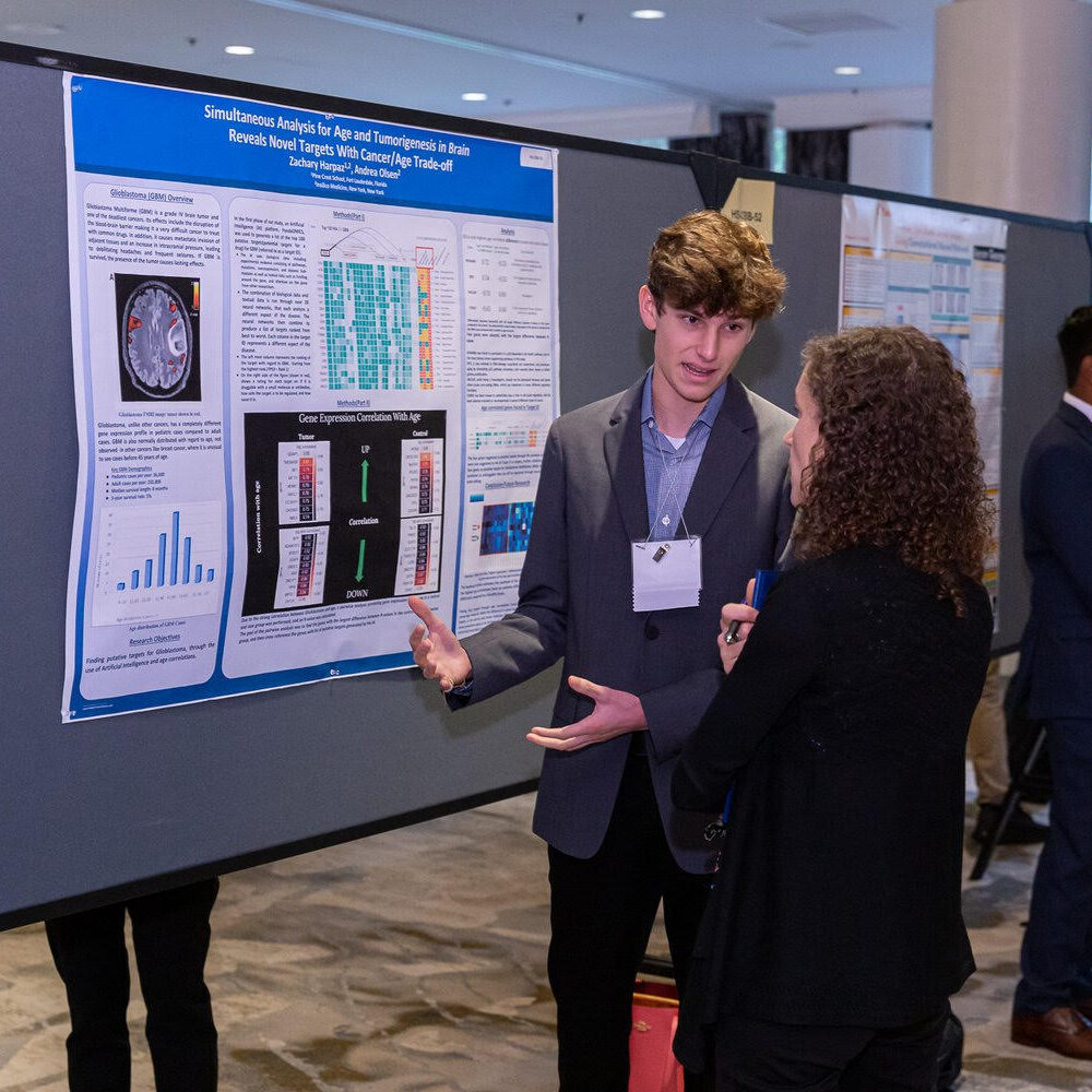
High School Students Use AI to Make Aging and Glioblastoma Discoveries

In a breakthrough study, three high school students and Insilico researchers used generative artificial intelligence (AI) to help identify new therapeutic targets for glioblastoma multiforme (GBM) and aging.

High School Students Use AI to Make Aging and Glioblastoma Discoveries
Listen to an audio version of this article

Glioblastoma multiforme (GBM) is one of the most aggressive and fatal malignant brain tumors. With a median survival time of 15 months, only about 25% of patients survive for one year and less than 5% survive for five years. As people get older, the risk of developing GBM increases. The discovery of new drug targets for GBM is of paramount importance.

The good news here is that high school students, Zachary Harpaz, Andrea Olsen and Christopher Ren, and researchers Anastasia Shneyderman, Alexander Veviorskiy, Maria Dralkina, Simon Konnov, Olga Shcheglova, Frank W. Pun, Geoffrey Ho Duen Leung, Hoi Wing Leung, Ivan V. Ozerov, Alex Aliper, Mikhail Korzinkin, and Alex Zhavoronkov have recently made remarkable strides in the joint field of aging and glioblastoma research. The team used a generative artificial intelligence (AI) engine from Insilico Medicine (founded by Dr. Alex Zhavoronkov) called PandaOmics, to identify new therapeutic targets for both GBM and aging. On April 26, 2023, their research paper was published in Aging’s Volume 15, Issue 8, entitled, “Identification of dual-purpose therapeutic targets implicated in aging and glioblastoma multiforme using PandaOmics – an AI-enabled biological target discovery platform.”

Their Study

Andrea Olsen, a student at Sevenoaks School in Kent, UK, and CEO/co-founder of The Youth Longevity Association
Photo of Andrea Olsen, courtesy of Insilico Medicine

“[Glioblastoma multiforme] is one of the most horrible cancers because it has such a short survival time,” Andrea Olsen said. “Of course, it affects the brain and so affects the body because the brain is the control center of the entire body.”

Andrea Olsen, a student at Sevenoaks School in Kent, UK, and CEO/co-founder of The Youth Longevity Association, discovered her interest in neurobiology and technology while growing up in Oslo, Norway. In 2021, she started an internship at Insilico Medicine. Through her work with the researchers at Insilico Medicine, Olsen learned how to use AI to uncover new genetic targets that could be used to treat aging and cancer. Zachary Harpaz, a student at Pine Crest School in Fort Lauderdale, Florida, discovered his passion for biology after being introduced to the subject in 2020. He combined his passion for biology with his intrigue for computer science and AI to enter the field of aging research.

Zachary Harpaz, a student at Pine Crest School in Fort Lauderdale, Florida
Photo of Zachary Harpaz, courtesy of Insilico Medicine

“We wanted to find new putative targets for glioblastoma as well as aging—attacking them both at the same time,” explained Harpaz.

The researchers used a comprehensive approach to identify their targets. They split their data into three categories—young, middle-aged and senior—and mapped the importance of gene expression to survival. They analyzed 12 datasets and selected the genes that were overlapped in 11 of the 12 datasets. They also cross-referenced those genes with a recent study conducted by the researchers at Insilico Medicine in 2022 on putative targets for aging and certain diseases. 

PandaOmics

One of the most exciting aspects of their research was the use of PandaOmics. Typically, finding new drugs requires experts to comb through a myriad of data and conduct extensive research. With PandaOmics, AI quickly processes and analyzes the data to identify new therapeutic targets, reducing the time and resources required for drug development. These high school researchers used PandaOmics to screen datasets from the Gene Expression Omnibus repository (maintained by the National Center for Biotechnology Information) and discovered three new potential therapeutic targets for treating both aging and GBM.

The first target for glioblastoma and aging was CNGA3, which they selected after analyzing each gene through PandaOmics. They also analyzed negatively correlated genes and selected GLUD1 as the number one gene produced by PandaOmics. Finally, they cross-referenced genes highly correlated to aging with the previous 2022 study and selected SIRT1 as another potential therapeutic target. The students were excited by the findings. Before she interned with Insilico, Olsen said she didn’t realize that AI could be so helpful in finding completely new therapeutic targets. 

“For me, [working with Insilico] was an incredible opportunity to dive into the field of research, aging, longevity, and neuroscience. It really kick-started my entire career,” Olsen said.

Looking Ahead

Overall, the study conducted by the researchers and these high school students showcases the power of AI in drug discovery and highlights the potential for young researchers to make meaningful contributions to the field. Their findings could lead to the development of new therapies for glioblastoma and aging-related diseases. Other therapeutics identified through Insilico’s Pharma.AI platform—specifically for idiopathic pulmonary fibrosis and COVID-19—have already advanced to human clinical trials.

“I’m excited to continue my research into college, but I’m super grateful for this opportunity at Insilico. It allowed me to get a head start on learning how to conduct research, analyze data and use the coolest and most cutting edge AI in the drug development area,” Harpaz said. “That experience gave me an amazing head start and I’m super excited to continue that into college, and even after college.”

Click here to read the full research paper published by Aging.

AGING (AGING-US) VIDEOS: YouTube | LabTube | Aging-US.com

Aging is an open-access, peer-reviewed journal that has been publishing high-impact papers in all fields of aging research since 2009. These papers are available to readers (at no cost and free of subscription barriers) in bi-monthly issues at Aging-US.com.

Click here to subscribe to Aging publication updates.

For media inquiries, please contact [email protected].

RNA Virus Fruit Fly Model: First Study to Measure Single-Fly Respiration

In a new study, researchers investigated the mortality and respiration rates of RNA virus-infected male fruit flies and how aging impacts these outcomes and measurements.

RNA Virus Fruit Fly Model: First Study to Measure Single-Fly Respiration

RNA viruses are responsible for approximately 70% of emerging infectious diseases in humans, according to a 2020 report by the National Academy of Medicine. Examples of RNA viruses include: influenza, hepatitis C, HIV, measles, zika, ebola, poliovirus, rhinovirus, rabies, and SARS-CoV-2—the virus responsible for the COVID-19 pandemic. After infection with an RNA virus, significant changes can take place in the host’s metabolism. While it is clear that disease tolerance declines as humans age, it is not yet clear how aging affects virus-induced changes in metabolism.

“Virus-induced metabolic reprogramming could impact infection outcomes, however, how this is affected by aging and impacts organismal survival remains poorly understood.”

In a new study, researchers Eli Hagedorn, Dean Bunnell, Beate Henschel, Daniel L. Smith Jr., Stephanie Dickinson, Andrew W. Brown, Maria De Luca, Ashley N. Turner, and Stanislava Chtarbanova from the University of Alabama, Indiana University, University of Arkansas for Medical Sciences, Arkansas Children’s Research Institute, and Jacksonville State University examined how an RNA virus can affect the respiration rate in male fruit flies (Drosophila melanogaster), both young and old. On March 22, 2023, their research paper was published in Aging’s Volume 15, Issue 6, entitled, “RNA virus-mediated changes in organismal oxygen consumption rate in young and old Drosophila melanogaster males.”

The Study

An organism’s metabolism depends on oxygen to produce energy. An efficient immune system depends, in part, on energy from the body’s metabolism to fuel it. Paradoxically, decreased metabolism, or hypometabolism, is a survival strategy that promotes disease tolerance in response to infection. In this study, the researchers used oxygen consumption rate (OCR) to indirectly measure changes in metabolism before and after RNA viral infection. The team infected male fruit flies with the RNA virus Flock House virus (FHV), and documented their oxygen consumption rate and/or mortality times at different time intervals after infection.

“As the exact mechanisms by which hypometabolism promotes tolerance are not fully understood, D. melanogaster could serve as an excellent model to dissect the genetic and molecular bases of this process.”

After the first 72-hours post-infection, FHV appeared to modulate respiration in all flies, but age did not appear to have a significant effect on OCR. However, over the course of the three-day experiment, the longitudinal assessment showed that OCR in young flies progressively and significantly decreased, while OCR in aged flies remained constant. The researchers found that the OCR at 24-hours varied in response to both experimental treatment and survival status. FHV-injected flies that died prior to 48- or 72-hours had a lower OCR compared to survivors at 48-hours. 

“Our results show that FHV infection significantly reduces organismal OCR compared to Tris-injected controls; however, we did not observe a significant change in OCR with aging. Interestingly, flies that died prior to 48-hours post-treatment measurements exhibited a significantly lower OCR at 24 h post-treatment compared to survivors. These findings suggest that the host’s metabolic profile could influence the outcome of viral infections.”

Conclusion

In conclusion, RNA viruses pose a significant threat to human health, causing numerous emerging infectious diseases. The impact of these viruses on the host’s metabolism, particularly in relation to aging, remains poorly understood. The recent study by Hagedorn et al. sheds light on the interaction between RNA viruses, metabolism and aging by examining the effects of the Flock House virus on the respiration rate of male fruit flies. The findings suggest that this infection can modulate the host’s OCR, and that the metabolic profile of the host could influence the outcome of viral infections.  The authors suggest that further research is needed to determine the precise mechanisms by which RNA viruses affect metabolic rate and to explore the potential for interventions to modulate metabolic rate and improve healthspan and lifespan.

“Older flies exhibit impaired disease tolerance to FHV [19], and here we show that metabolic rate depression does not occur in older flies in response to FHV in the first three days following treatment. It is therefore possible that as is the case in mammals, flies employ hypometabolism as a survival strategy that is part of a disease tolerance mechanism. It would be interesting in the future to test this hypothesis by comparing OCR in tolerance mutant flies such as the G9a mutants.”

Click here to read the full research paper published by Aging.

AGING (AGING-US) VIDEOS: YouTube | LabTube | Aging-US.com

Aging is an open-access, peer-reviewed journal that has been publishing high-impact papers in all fields of aging research since 2009. These papers are available to readers (at no cost and free of subscription barriers) in bi-monthly issues at Aging-US.com.

Click here to subscribe to Aging publication updates.

For media inquiries, please contact [email protected].

A Promising Approach to Preventing Periodontitis

A new study by researchers from Osaka University’s Graduate School of Dentistry investigated cellular senescence in periodontal tissue and disease—identifying promising therapeutic targets for preventing periodontitis in the elderly.

A new study by researchers from Osaka University’s Graduate School of Dentistry investigated cellular senescence in periodontal tissue and disease—identifying promising therapeutic targets for preventing periodontitis in the elderly.

The Trending With Impact series highlights Aging publications (listed by MEDLINE/PubMed as “Aging (Albany NY)” and “Aging-US” by Web of Science) that attract higher visibility among readers around the world online, in the news and on social media—beyond normal readership levels. Look for future science news about the latest trending publications here, and at Aging-US.com.

Listen to an audio version of this article

Repercussions of poor dental health aren’t limited to mere social stigmas. Poor dental health can impart serious consequences on an individual’s overall health. Periodontal disease broadly refers to any disease that affects the gums and the surrounding tissues that support the teeth, including the periodontal ligament (PDL) and alveolar bone. Periodontal disease can increase the risk of heart disease, stroke and diabetes by allowing bacteria to enter the bloodstream, causing inflammation and organ damage. 

Periodontitis is a more advanced stage of periodontal disease. It is thought to be the most common infectious disease in the United States—affecting more than 40% of adults over 30 years old. Previous research has suggested that aging is a significant risk factor for periodontitis, although the underlying mechanisms are unclear.

“The direct cause of periodontitis is periodontopathic bacteria, while various environmental factors affect the severity of periodontitis. Previous epidemiological studies have shown positive correlations between aging and periodontitis. However, whether and how aging is linked to periodontal health and disease in biological processes is poorly understood.”

In a recent study, researchers Kuniko Ikegami, Motozo Yamashita, Mio Suzuki, Tomomi Nakamura, Koki Hashimoto, Jirouta Kitagaki, Manabu Yanagita, Masahiro Kitamura, and Shinya Murakami from Osaka University’s Graduate School of Dentistry aimed to elucidate the underlying mechanisms that contribute to aging-associated inflammation in periodontitis. On March 1, 2023, their new research paper was published in Aging (Aging-US) Volume 15, Issue 5, entitled, “Cellular senescence with SASP in periodontal ligament cells triggers inflammation in aging periodontal tissue.”

The Study

“In this study, we aimed to clarify the pathophysiological roles of cellular senescence in periodontal tissue and diseases.”

Previous studies have found that senescent cells can secrete senescence-associated secretory proteins (SASP) that induce inflammation and impair wound healing in some chronic diseases. The existence of senescent cells in periodontal tissue and diseases, however, has yet to be clarified. In this study, the researchers investigated cellular senescence and SASP in aging periodontal tissue. The team aimed to uncover the mechanism by which cellular senescence and SASP trigger inflammation in periodontal tissue and to identify potential therapeutic targets for this disease. 

To investigate the role of cellular senescence in periodontitis, the researchers analyzed periodontal tissue in young and aged mice. Alveolar bone volume was compared in the young and aged mice, and beta-galactosidase (β-gal) staining was performed. They found bone resorption in aged mice and many senescence-associated (SA) β-gal-positive cells in their periodontal tissue, leading to inflammation and breakdown of alveolar bone. Very few SA β-gal-positive cells were found in young mouse tissues.

Next, the researchers worked with cells in vitro, primary human periodontal ligament (HPDL) cells, and induced cellular senescence through serial passaging (replicative senescence). The growth rate of HPDL cells gradually reduced, and they reached irreversible cell growth arrest, indicating the induction of cellular senescence. Morphological changes were observed through phalloidin staining. The team found that around 70% of aged HPDL cells were positive for SA β-gal, while less than 10% of young HPDL cells were positive. Morphological changes showed that aged HPDL cells had an enlarged and “spread” cell shape compared to young HPDL cells. Flow cytometry analysis confirmed an increase in cell size and granularity of aged HPDL cells compared to young HPDL cells.

TEM analysis showed that senescent cells exhibit metabolic changes and irregularly shaped mitochondria with disrupted cristae and increased accumulation of ROS, which suggest damage and failure of the redox balance. Importantly, the researchers found that the intrinsic inflammation state of aged PDLs was higher than in young PDLs, and susceptibility to bactericidal pathogens (but not inflammatory cytokines) was low in aged PDLs. Additionally, the team observed an age-dependent upregulation of microRNA (miR)-34a in HPDL cells.

“Thus, miR-34a and senescent PDL cells might be promising therapeutic targets for periodontitis in elderly people.”

Summary & Conclusion

In conclusion, poor dental health can have serious implications on an individual’s overall health, as periodontal disease can increase the risk of heart disease, stroke and diabetes. Aging is a significant risk factor for periodontitis, which is thought to be the most common infectious disease in the United States. A recent study by researchers from Osaka University’s Graduate School of Dentistry aimed to elucidate the underlying mechanisms that contribute to aging-associated inflammation in periodontitis. The study found that senescent cells in periodontal tissue secrete SASP that induce inflammation. The researchers identified potential therapeutic targets for periodontitis and suggest that elimination of senescent PDL cells or suppression of the miR-34a-dependent SIRT1-NF-κB axis may be an attractive therapeutic strategy to prevent periodontitis in humans as we age.

“To the best of our knowledge, this is the first study to identify: 1) the potential for senescent PDL cells to induce inflammation of periodontal tissue, and 2) a miRNA-dependent molecular mechanism of SASP in senescent PDL cells.”

Click here to read the full research paper published by Aging.

AGING (AGING-US) VIDEOS: YouTube | LabTube | Aging-US.com

Aging is an open-access, peer-reviewed journal that has been publishing high-impact papers in all fields of aging research since 2009. These papers are available to readers (at no cost and free of subscription barriers) in bi-monthly issues at Aging-US.com.

Click here to subscribe to Aging publication updates.

  • Follow Us