Aging’s Commitment to Advancing Research: Sponsoring the “Future of Aging Research Mixer 2024”

Future of Aging Research Mixer

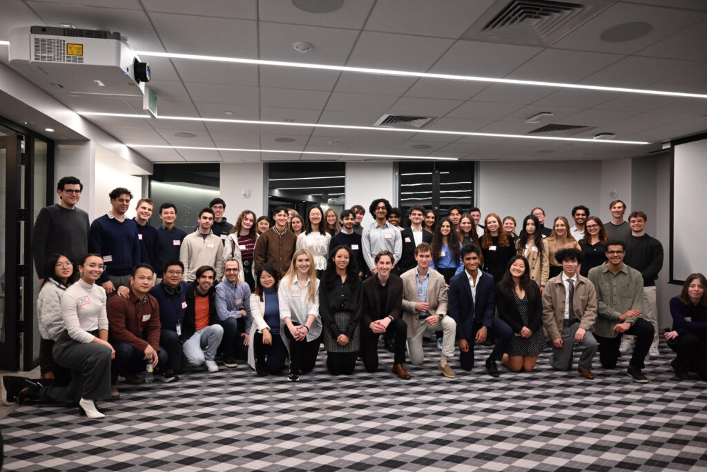
Aging (Aging-US) was a proud sponsor of the “Future of Aging Research Mixer 2024” hosted by the Aging Initiative at Harvard University on November 15 in Boston. This event united a vibrant community of students, researchers and technologists, all driven by a shared mission: advancing innovations in aging research and longevity science.

Key Highlights from the Future of Aging Research Mixer 2024

The event kicked off with inspiring opening remarks and a keynote by George Church, professor at Harvard Medical School, founding member of the Wyss Institute, and co-founder of over 50 biotech companies. He was joined by Kat Kajderowicz, an MIT PhD student and Principal at age1. Together, they highlighted the interdisciplinary nature of aging research and its immense potential to drive transformative advancements.

Jesse Poganik, HMS Instructor in Medicine and Executive Co-Director of the Biomarkers of Aging Consortium, discussed the evolution of aging science and the critical role biomarkers play in understanding aging processes and assessing the effectiveness of interventions aimed at slowing or reversing age-related changes.

Alex Colville, co-founder and general partner at age1, explained how venture capital can accelerate innovation in longevity biotechnology. He shared career advice for aspiring researchers and paid tribute to his mentor, Dr. David Sinclair, a pioneer in aging research.

These talks highlighted the importance of mentorship, interdisciplinary collaboration, and investment in driving progress in the aging research field.

Empowering Future Aging Science Leaders

A majority of the attendees were students from Boston-area universities including Harvard, MIT, UMass and BU. These future scientists, entrepreneurs, and innovators engaged in meaningful discussions about research, career paths, and publishing in academic journals. Many expressed interest in journals like Aging (Aging-US) and sought advice on how to publish their work.

The “Future of Aging Research Mixer 2024” showcased the passion, collaboration, and innovation within the aging research community. Through its sponsorship, Aging (Aging-US) reaffirmed its commitment to fostering a vibrant network of talent and supporting the voices of young, passionate researchers. Initiatives like this inspire the next generation of scientists and entrepreneurs, driving sustained growth and transformative impact in the field.

Beyond the event, the Aging Initiative at Harvard University strengthens the community through ongoing programs like journal clubs, guest lectures, and informal lunches with professors. These initiatives encourage skill-building, idea-sharing, and mentorship, preparing students for impactful careers in aging science.

Why We Support Aging Research

Aging (Aging-US) was founded in 2008 by visionary scientists—the late Dr. Mikhail (Misha) Blagosklonny, the late Dr. Judith Campisi, and Dr. David Sinclair—with a clear mission: to create a journal by scientists, for scientists, so the researchers can publish their ideas, theories (sometimes unconventional) and studies on the rapidly developing aging field. Since then, we have remained dedicated to advancing the understanding of aging and age-related diseases, including cancer, a leading health challenge in today’s aging world.

Supporting initiatives like the Aging Initiative at Harvard University and events such as the “Future of Aging Research Mixer 2024” is central to our mission. By supporting young researchers, we strive to drive meaningful advancements in the field and ensure it receives the recognition and resources it deserves. We are deeply committed to supporting initiatives that empower scientists and promote collaboration, mentorship, and innovation.

Sponsoring this initiative is more than an investment—it’s a commitment to the future of aging science and a healthier, longer life for all.

As we look to the future, we are inspired by the passion and talent within this growing field. Together, through continued collaboration and investment, we can shape a world where aging research leads to healthier and longer lives.

Aging is indexed by PubMed/Medline (abbreviated as “Aging (Albany NY)”), PubMed CentralWeb of Science: Science Citation Index Expanded (abbreviated as “Aging‐US” and listed in the Cell Biology and Geriatrics & Gerontology categories), Scopus (abbreviated as “Aging” and listed in the Cell Biology and Aging categories), Biological Abstracts, BIOSIS Previews, EMBASE, META (Chan Zuckerberg Initiative) (2018-2022), and Dimensions (Digital Science).

Click here to subscribe to Aging publication updates.

For media inquiries, please contact [email protected].
How AI and Longevity Biotechnology are Revolutionizing Healthcare for Healthier, Longer Lives

“The integration of artificial intelligence (AI), biomarkers, ageing biology, and longevity medicine stands as a cornerstone for extending human healthy lifespan.”

Imagine a future where we not only live longer but stay healthy throughout those extra years. Thanks to recent breakthroughs in biotechnology and artificial intelligence (AI) in healthcare, this vision is closer to becoming a reality.

Advancements in Aging Research

Aging research has made significant progress in recent years by combining disciplines like biology, technology, and medicine to tackle the challenges of extending healthspans and reducing age-related diseases. While people today live longer than ever before, extending our “healthspan”—the years we stay active and illness-free—remains challenging. AI and health biomarkers (biological indicators of our body’s condition) are now key tools in the pursuit of longer, healthier lives.

In a recent paper, led by corresponding authors Yu-Xuan Lyu from Southern University of Science and Technology Shenzhen; Alex Zhavoronkov from Insilico Medicine AI Limited, Masdar City, Abu Dhabi; Morten Scheibye-Knudsen and Daniela Bakula from the Center for Healthy Aging, University of Copenhagen, along with numerous other collaborators, the transformative potential of AI in aging research was explored. The research paper, titled “Longevity biotechnology: bridging AI, biomarkers, geroscience and clinical applications for healthy longevity,” was published as the cover paper in Aging’s Volume 16, Issue 20.

The Study: A New AI-Powered Approach to Aging

The work summarizes insights from the 2023 Aging Research and Drug Discovery Meeting. Researchers from renowned institutions explored how AI, biomarkers, and clinical applications can work together to enhance longevity. This fusion, termed “longevity biotechnology,” promises to transform healthcare from reactive treatments to proactive, preventive measures focused on staying healthy as we age.

The Challenge: Targeting Multiple Health Conditions with Longevity Biotechnology

Traditional aging research often targets single diseases, but most elderly individuals experience multiple chronic conditions. Addressing this complex challenge requires identifying biological markers that indicate aging and predicting health risks before diseases manifest.

The Breakthrough: AI in Biomarker Discovery for Aging

The study highlights how AI can accelerate the discovery of biomarkers, allowing scientists to understand aging at the cellular level. By using machine learning to identify unique patterns, researchers can estimate biological age, discover potential treatments, and evaluate the impact of lifestyle changes on health. This personalized approach enables healthcare providers to create prevention and treatment plans suited to each person’s unique health needs.

The Future of Healthcare: Preventive, AI-Driven Longevity Treatments

Currently, healthcare often focuses on managing diseases as they arise. However, these AI-driven tools could bring about a shift to preventive healthcare. Instead of waiting for age-related illnesses, clinicians could use AI insights to address aging’s root causes, improving health before issues arise.

While the promise of AI in healthcare is significant, the research team emphasizes that further investment is needed to make these AI-driven approaches accessible and accurate. With continued advancements, longevity biotechnology could become a standard part of healthcare, offering a new way to maintain vitality and well-being as we age.

Conclusion

Longevity biotechnology represents a groundbreaking shift, with AI and biomarkers helping us envision a future of healthier, longer lives. This approach brings us closer to understanding and managing the aging process, making extended healthspans a real possibility.

Click here to read the full research paper in Aging.

Aging is indexed by PubMed/Medline (abbreviated as “Aging (Albany NY)”), PubMed CentralWeb of Science: Science Citation Index Expanded (abbreviated as “Aging‐US” and listed in the Cell Biology and Geriatrics & Gerontology categories), Scopus (abbreviated as “Aging” and listed in the Cell Biology and Aging categories), Biological Abstracts, BIOSIS Previews, EMBASE, META (Chan Zuckerberg Initiative) (2018-2022), and Dimensions (Digital Science).

Click here to subscribe to Aging publication updates.

For media inquiries, please contact [email protected].

How Single Housing Impacts Growth and Lifespan in African Turquoise Killifish

“[…] our results suggest that sharing housing with others in early life might influence whole-life attributes, potentially leading to specific life history traits beyond the typical relationship between the growth rate and lifespan.”

In this research, Chika Takahashi, Emiko Okabe, Masanori Nono, Saya Kishimoto, Hideaki Matsui, Tohru Ishitani, Takuya Yamamoto, Masaharu Uno, and Eisuke Nishida from the RIKEN Center for Biosystems Dynamics Research (BDR) in Hyogo, Japan; Brain Research Institute, Niigata University in Niigata, Japan; Research Institute for Microbial Diseases at Osaka University in Osaka, Japan; Kyoto University in Kyoto, Japan; and RIKEN Center for Advanced Intelligence Project (AIP), explored the effects of housing density during the juvenile stage on whole-life traits, including growth, fecundity, and lifespan, in African turquoise killifish. Their research paper was published on the cover of Aging (listed by MEDLINE/PubMed as Aging (Albany NY) and as Aging-US by Web of Science), Volume 16, Issue 18, entitled, “Single housing of juveniles accelerates early-stage growth but extends adult lifespan in African turquoise killifish.”

THE STUDY

A study on African turquoise killifish examined the impact of housing density on juvenile growth. Newly hatched fish were kept in different densities ranging from 1 to 40 fish per tank. It was found that lower housing densities resulted in faster growth, with fish in single housing growing significantly larger than those in group housing. Additionally, single-housed fish reached sexual maturity earlier compared to group-housed fish at higher densities. Comparisons between group-housed and single-housed fish showed that housing conditions in the juvenile stage did not affect the appearance changes during sexual maturation. 

As the fish progressed to middle-aged adults, the rate of increase in body length slowed down, while body weight continued to increase. Differences in body weight between group-housed and single-housed fish persisted into old age, suggesting potential differences in body composition. Surprisingly, single-housed fish had a longer mean adult lifespan compared to group-housed fish, contradicting the commonly held belief that faster growth leads to shorter lifespan. Lower housing densities during the juvenile stage were also found to extend adult lifespan, further challenging the inverse correlation between growth rate and lifespan. These findings suggest that lower housing densities promote accelerated growth in the juvenile stage of African turquoise killifish.

The study also found that single-housed fish had a longer adult lifespan compared to group-housed fish. This led to the suspicion that the egg-laying period of single-housed fish might also be longer. To investigate this, the researchers conducted weekly monitoring of the number of eggs laid until the old adult stage. In group-housed fish, the number of eggs laid was high for the first two weeks, followed by a medium level for the subsequent five weeks, and then decreased. In contrast, single-housed fish showed a medium level of egg-laying for the first nine weeks, followed by a decrease. The cumulative number of live embryos was found to be lower in single-housed fish compared to group-housed fish. These findings suggest that while the number of eggs laid is not very high, single-housed fish have a longer egg-laying period than group-housed fish.

To investigate the potential reasons behind the reduction in offspring number and longer egg-laying period in single-housed fish, the researchers conducted RNA sequencing analysis of testes or ovaries at four life stages. These stages included the onset of sexual maturity, young adult, mature adult, and middle-aged adult. Interestingly, the analysis revealed that single-housed fish showed higher similarity to group-housed fish at earlier life stages compared to group-housed fish at the same life stage. For instance, in the testes, single-housed fish at stage II exhibited the highest similarity to group-housed fish at stage I. Similarly, in the ovaries, single-housed fish at stage II and III showed higher similarity to group-housed fish at stage I. These findings suggest that the rate of gonadal transcriptional change with life stage progression is slower in single-housed fish compared to group-housed fish.

The researchers identified differentially expressed genes (DEGs) between stage I and stage IV in group- and single-housed fish. In the testes, ribosome-related genes and cilium-related genes were highly enriched in DEGs with higher expression in stage I compared to stage IV, suggesting a link between life stage progression, testes development, and spermatogenesis. In the ovaries, growth-related genes and translation-related genes were highly enriched in DEGs with higher expression in stage I compared to stage IV, indicating a link between life stage progression, ovarian development, oogenesis, and aging. Comparing group-housed and single-housed fish at different stages, there were differences in the PC1 values, suggesting that single-housed fish exhibited slower progression of gametogenesis and gonadal maturation relative to life stage progression compared to group-housed fish.

To further investigate this, the researchers focused on specific genes related to spermatogenic differentiation, oocyte development, oocyte construction, and female gonad development. The expression of these genes showed slower changes with life stage progression in single-housed fish compared to group-housed fish in both the testes and ovaries. This suggests that single-housed fish may have slower rates of gametogenesis and gonadal maturation, leading to a lower proportion of mature sperm and oocytes in their gonads. Overall, the results indicate that, at the transcriptional level, the progression of gonadal maturation and ovarian aging is slower in single-housed fish compared to group-housed fish. This slower progression may explain the medium fecundity and extended egg-laying period observed in single-housed fish.

The liver was chosen for analysis as it plays a central role in organismal metabolic processes. Gene expression profiles of the livers were compared between group- and single-housed fish at two different ages: 7 weeks post-hatching (wph) and 14 wph. Surprisingly, despite the 2-week age difference, the correlation coefficients showed that group- and single-housed fish at 14 wph were highly similar. The researchers identified 1588 age-related differentially expressed genes (DEGs) between the two age groups. Hierarchical clustering based on the expression changes of these age-related genes demonstrated that the expression profiles of group- and single-housed fish were similar at 14 wph.

IN CONCLUSION

In summary, juvenile single housing in African turquoise killifish promotes faster growth, longer egg-laying periods, and extended lifespans compared to group housing. These findings challenge traditional assumptions about the relationship between growth and lifespan and shed light on the impact of early-life environmental conditions on overall life history.

Overall, the experiments involved maintaining and rearing the fish, measuring their body length and weight, analyzing RNA sequencing data, measuring lifespan, and counting the number of eggs laid. Statistical analysis was conducted to assess significant differences between groups.

Click here to read the full research paper in Aging.

Aging is indexed by PubMed/Medline (abbreviated as “Aging (Albany NY)”), PubMed CentralWeb of Science: Science Citation Index Expanded (abbreviated as “Aging‐US” and listed in the Cell Biology and Geriatrics & Gerontology categories), Scopus (abbreviated as “Aging” and listed in the Cell Biology and Aging categories), Biological Abstracts, BIOSIS Previews, EMBASE, META (Chan Zuckerberg Initiative) (2018-2022), and Dimensions (Digital Science).

Click here to subscribe to Aging publication updates.

For media inquiries, please contact [email protected].

The Cell Rejuvenation Atlas: Unveiling Rejuvenation Strategies through Network Biology

Researchers introduce SINGULAR, a cell rejuvenation atlas that provides a unified analysis framework to study the effects of rejuvenation strategies at the single-cell level.

Researchers Javier Arcos Hodar, Sascha Jung, Mohamed Soudy, Sybille Barvaux, and Antonio del Sol from CIC bioGUNE-BRTA and University of Luxembourg introduce SINGULAR, a cell rejuvenation atlas that provides a unified analysis framework to study the effects of rejuvenation strategies at the single-cell level. On September 9, 2024, their research paper was published on the cover of Aging (listed by MEDLINE/PubMed as “Aging (Albany NY)” and “Aging-US” by Web of Science), Volume 16, Issue 17, entitled, “The cell rejuvenation atlas: leveraging network biology to identify master regulators of rejuvenation strategies.”

THE RESEARCH

Various strategies, including lifestyle changes, gene therapies, and surgical procedures, have shown promise in improving aging markers and increasing lifespan in model organisms. These interventions often have limitations, however, such as not achieving comprehensive functional improvement across tissues or facing challenges in clinical translation. To address these limitations, the researchers characterized and compared rejuvenation interventions at different biological levels. The paper introduces SINGULAR, a cell rejuvenation atlas that provides a unified analysis framework to study the effects of rejuvenation strategies at the single-cell level. By examining gene regulatory networks, intracellular signaling, cell-cell communication, and cellular processes, the atlas identifies master regulators and common targets across immune cells. SINGULAR has the potential to inform future advancements in human age reversal and aid in the selection of drugs that mimic the effects of rejuvenation interventions.

RESULTS

The authors propose a unified multiscale analysis pipeline for characterizing and comparing the effects of rejuvenation interventions. This process begins by filtering low-quality cells, normalizing expression profiles, and identifying optimal cell clustering. The data is then analyzed at various biological levels, including differential gene expression, transcriptional regulatory networks, signaling cascades, and intercellular communication.

Nine previously published single-cell RNA-seq datasets from different rejuvenation interventions were collected and analyzed, revealing technical variability that highlights the need for a standardized data processing pipeline. The analysis showed heterogeneous gene expression responses across different cell types and organs. Systemic interventions had consistent effects on multiple organs, while metformin had minimal impact. Interestingly, exercise produced the largest transcriptional effects in the liver, artery, and spinal cord, even though it primarily targets muscles.

Transcriptional regulatory networks (TRNs) were reconstructed to explore the regulatory mechanisms behind these gene expression changes. The TRNs, which averaged 72 genes, were highly hierarchical, indicating the presence of ‘master regulators’ that explain significant portions of gene expression changes.

To demonstrate the practical application of SINGULAR, the study investigated the identification of drugs that could target transcription factor (TF) master regulators and key signaling molecules. Drug-target relationships from DrugBank were analyzed to find drugs that could activate master regulators or mimic the effects of rejuvenation interventions. Interestingly, only 17 out of 239 TFs could be activated by drugs, primarily nuclear receptors, with notable exceptions like AP-1 complex proteins and Trp53. Some of these drugs, such as Curcumin and Vitamin D3, have shown rejuvenating effects on lifespan in model organisms. Key signaling molecules were found to be more druggable, with several drugs targeting specific molecules, though none targeted both genes.

The study aimed to identify master regulators and their downstream effects in rejuvenation interventions. By simulating the activation of transcription factors (TFs) within the network, the researchers quantified the number of genes regulated by each TF. They discovered 493 TFs with non-zero activity across various conditions, though most acted as master regulators in only a few cases. The study also highlighted key differences between TFs involved in aging-related activity changes and those regulating rejuvenation. Notably, the AP-1 complex, consisting of Fos and Jun, emerged as a common master regulator across multiple interventions. The researchers also identified TFs linked to aging and validated their potential rejuvenating effects experimentally. They also explored crosstalk between TFs and signaling pathways, finding negative enrichment of aging gene sets in several integrated networks. Overall, the findings offer valuable insights into the regulatory mechanisms and potential rejuvenating effects of master regulators and signaling molecules involved in rejuvenation interventions.

CONCLUSION

In conclusion, this study employed a unified analysis pipeline, SINGULAR, to compare the effects and mediators of various rejuvenation interventions. Key master regulators, including Arntl, AP-1 complex proteins, NFE2L2, and MAF, were identified as playing crucial roles in rejuvenation. The analysis revealed distinct differences between aging-related transcriptional changes and rejuvenation regulators. Immune and skin cell types were highlighted as potential intervention targets, with the possibility of additive or synergistic effects by targeting non-overlapping master regulators. Some limitations were noted, such as biases in cell type comparisons, reliance on ligand-receptor interactions for cell-cell communication analysis, and the risk of false negatives in differential expression testing. Despite these limitations, SINGULAR offers valuable insights into rejuvenation mechanisms and the identification of agents for anti-aging strategies. It provides a robust framework for understanding the mechanisms behind various interventions and offers a wide range of potential target genes for a comprehensive anti-aging approach.

Click here to read the full research paper in Aging.

Aging is indexed by PubMed/Medline (abbreviated as “Aging (Albany NY)”), PubMed CentralWeb of Science: Science Citation Index Expanded (abbreviated as “Aging‐US” and listed in the Cell Biology and Geriatrics & Gerontology categories), Scopus (abbreviated as “Aging” and listed in the Cell Biology and Aging categories), Biological Abstracts, BIOSIS Previews, EMBASE, META (Chan Zuckerberg Initiative) (2018-2022), and Dimensions (Digital Science).

Click here to subscribe to Aging publication updates.

For media inquiries, please contact [email protected].

Unveiling Role of Cytoskeleton in Aging: Insights from Dermal Fibroblast Research

In this study, researchers reinforce knowledge about an age-related alteration in the synthesis of major proteins linked to the migratory and contractile functions of dermal human fibroblasts.

Dermal fibroblasts orchestrate the synthesis and degradation of extracellular matrix components, which is crucial for skin homeostasis. Alterations in the expression of components such as collagens and enzymes can lead to reduced mechanical cutaneous tension and impaired skin wound healing during aging.

Researchers Françoise Boismal, Sandy Peltier, Sophie Ly ka so, Guillaume Chevreux, Loïse Blondel, Kévin Serror, Niclas Setterblab, Elina Zuelgaray, David Boccara, Maurice Mimoun, Christelle Guere, Armand Benssussan, Marie Dorr, Gallic Beauchef, Katell Vie, and Laurence Michel from Saint-Louis Hospital, ParisParis University, Paris CitéJacques-Monod Institute, Paris; and Clarins Laboratories, Pontoise, aimed to better understand the molecular alterations in fibroblasts during aging by comparing secretomic and proteomic signatures of fibroblasts from young (<35years) and aged (>55years) skin donors, in quiescence or TGF-stimulated conditions, using HLPC/MS. 

Their research paper was published on the cover of Aging’s Volume 16, Issue 16, entitled, “Proteomic and secretomic comparison of young and aged dermal fibroblasts highlights cytoskeleton as a key component during aging.”

Dermal fibroblasts were obtained from healthy, sun-protected skin of young (<35 years) and aged (>55 years) healthy women undergoing breast reduction surgery. Peptides were loaded using an online preconcentration method and separated by chromatography. RNA extraction, reverse transcription, quantitative PCR, and blot quantification were performed, along with immunostaining on fibroblasts seeded on culture chamber slides.

To identify key molecules involved in the role of human dermal fibroblasts during wound healing and skin aging, a comparative analysis of the secretome and proteome of 12 fibroblast cultures, freshly isolated from young and mature skin, was conducted using HPLC/MS. This analysis was performed in both quiescence and TGF-β1-treated conditions, without senescence-inducing factors, as described in previously reported aging models. Importantly, the analyses were conducted in the absence of serum in the culture medium 24 hours before and during cell stimulation to avoid serum protein contamination in the secretomic and proteomic assays

This study revealed a significant decrease in fibroblast protein secretion with age, while cytoplasmic protein accumulation increased by over 60%. Proteins related to actin and ECM (extracellular matrix) organization were the two main categories altered during aging. An in-depth analysis of actin-related proteins highlighted the involvement of CFL1, CORO1C, the ARP2/3 complex, FLNB, and ACTC1 in cytoskeleton organization and fibroblast migration. These findings offer potential new targets to slow key features of skin aging.

“Our present data reinforce knowledge about an age-related alteration in the synthesis of major proteins linked to the migratory and contractile functions of dermal human fibroblasts.”

Read the full research paper, published in Aging.

Aging is an open-access, traditional, peer-reviewed journal that publishes high-impact papers in all fields of aging research. All papers are available to readers (at no cost and free of subscription barriers) in bi-monthly issues at Aging-US.com.

Click here to subscribe to Aging publication updates.

For media inquiries, please contact [email protected].

Links Between Exercise, Senescence, and Lung Health

In a new study, researchers investigated myocyte-secreted factors with the potential to suppress cellular senescence, aiming to explore their protective effects against lung disease.

Over the human lifespan, our cells encounter numerous stressors that can trigger an intrinsic defense mechanism called cellular senescence. Cellular senescence is characterized by irreversible growth arrest and can act as a safeguard against cancer. However, when senescent cells accumulate in various tissues as we age, it can contribute to tissue degeneration and chronic diseases. 

The senescence-associated secretory phenotype (SASP), a hallmark of senescent cells, plays a critical role by secreting inflammatory factors, proteases, and growth factors, disrupting tissue balance and fueling pathological conditions. Consequently, selectively eliminating senescent cells has emerged as a promising therapeutic strategy, potentially restoring tissue function and mitigating age-related disorders.

COPD: Chronic Obstructive Pulmonary Disease

Chronic obstructive pulmonary disease (COPD) exemplifies the impact of cellular senescence on health, characterized by the collapse of alveolar walls in the lungs. Accelerated accumulation of senescent cells in COPD patients’ lung tissues links senescence to the disease’s pathogenesis. Genetic or pharmacological elimination of these cells in preclinical models has shown significant reductions in emphysema-associated pathologies and restoration of pulmonary function, highlighting the potential of senolytic therapies.

Regular physical activity offers benefits beyond fitness, including cardiovascular and mental well-being enhancements, and modulates cellular senescence. Studies show an association between habitual exercise and lower levels of senescence markers in various tissues. Researchers have focused on myokines, signaling factors secreted by skeletal muscles in response to exercise, as potential mediators of these benefits. Irisin, a myokine, has shown promise in suppressing cellular senescence and correlating inversely with COPD severity.

In a new study, researchers Hiromichi Tsushima, Hirobumi Tada, Azusa Asai, Mikako Hirose, Tohru Hosoyama, Atsushi Watanabe, Taro Murakami, and Masataka Sugimoto from Tokyo Metropolitan Institute for Geriatrics and Gerontology, Shigakkan University, and National Center for Geriatrics and Gerontology investigated myocyte-secreted factors with the potential to suppress cellular senescence, aiming to explore their protective effects against lung disease. Their research paper was published on the cover of Aging’s Volume 16, Issue 13, entitled, “Roles of pigment epithelium-derived factor in exercise-induced suppression of senescence and its impact on lung pathology in mice.”

PEDF: A Promising Senescence Suppressor

In this recent study, pigment epithelium-derived factor (PEDF) emerged as a key player in the interplay between exercise, cellular senescence, and lung pathologies. Initially known for its role in retinal development, PEDF has been linked to cellular senescence modulation, extending the replicative lifespan of fibroblasts and diminishing senescence markers. PEDF mitigates oxidative stress by reducing reactive oxygen species levels and modulates microRNAs, particularly miR-127, implicated in cellular senescence.

“We found that myocyte-derived factors significantly extended the replicative lifespan of fibroblasts, suggesting that myokines mediate the anti-senescence effects of exercise.”

Exercise significantly upregulates PEDF expression in skeletal muscles, correlating with reduced senescence markers and SASP-related genes in the lungs. Recombinant PEDF administration in mice has shown remarkable results, reducing senescence markers and preserving alveolar structure in pulmonary emphysema models, translating into improved pulmonary function. While some preclinical evidence supports PEDF’s therapeutic potential, translating these findings to clinical applications requires rigorous safety and efficacy evaluations. Understanding PEDF’s signaling pathways could unveil new therapeutic targets, and its potential involvement in other age-related disorders warrants further investigation. The interplay between PEDF and other exercise-induced factors offers potential for novel therapeutic strategies.

“Collectively, these results strongly suggest that PEDF contributes to the beneficial effects of exercise, potentially suppressing cellular senescence and its associated pathologies.”

Conclusions

The discovery of PEDF’s role in exercise-induced senescence suppression and its therapeutic potential in lung pathologies represents a paradigm shift in senescence research. Understanding the interplay between physical activity, myokine signaling, and senescence modulation can lead to targeted interventions promoting healthy aging. Multidisciplinary collaborations are essential to harness the potential of PEDF and other senescence-modulating factors, paving the way for innovative treatments that alleviate age-related diseases and improve quality of life.

Read the full research paper, published in Aging.

Aging is an open-access, traditional, peer-reviewed journal that publishes high-impact papers in all fields of aging research. All papers are available to readers (at no cost and free of subscription barriers) in bi-monthly issues at Aging-US.com.

Click here to subscribe to Aging publication updates.

For media inquiries, please contact [email protected].

Targeting Mitophagy as a Therapeutic Strategy for Cancer Treatment

In this new study, researchers investigated the intricate link between mitophagy and cancer stem cells. 

Cellular quality control mechanisms like mitophagy, a specialized form of autophagy that eliminates dysfunctional mitochondria, play a pivotal role in various physiological processes. Defects in mitophagy have been linked to neurodegeneration, heart failure, cancer, and aging.

A recent study, by researchers Marta Mauro-Lizcano, Federica Sotgia, and Michael P. Lisanti from the University of Salford, has shed light on the intricate link between mitophagy and cancer stem cells (CSCs). In this study, the researchers developed an innovative fluorescence-based approach to enrich subpopulations of cancer cells exhibiting high basal levels of mitophagy. Their findings reveal that elevated mitophagy activity enhances CSC properties, including self-renewal, ATP production, proliferation, and cell migration, underscoring the potential of targeting mitophagy as a therapeutic strategy for cancer treatment. 

On June 4, 2024, their research paper was published on the cover of Aging’s Volume 16, Issue 11, entitled, “Mitophagy and cancer: role of BNIP3/BNIP3L as energetic drivers of stemness features, ATP production, proliferation, and cell migration.”

“CSCs are responsible for cancer relapse, therapy-resistance, and metastatic dissemination. Therefore, CSC elimination is necessary to prevent cancer recurrence and improve long-term patient outcomes. The search of new targets against CSCs is essential for the success of cancer treatment.” — Mauro-Lizcano et al.

Background

Mitophagy plays a crucial role in maintaining cellular homeostasis by selectively degrading damaged or superfluous mitochondria. This process is governed by specific mitochondrial outer membrane receptors, such as BNIP3 and BNIP3L (also known as NIX), which interact with autophagy-related proteins like LC3/GABARAP to initiate mitophagy. The current study focused on the BNIP3/BNIP3L-dependent pathway, which is rapidly induced under cellular stress conditions like hypoxia and nutrient deprivation.

Cancer stem cells (CSCs) are a subpopulation of cells within a tumor that exhibit stem cell-like properties, such as self-renewal, tumor initiation capability, and drug resistance. These cells are implicated in cancer recurrence, treatment failure, and metastatic dissemination, making their elimination a critical target for effective cancer therapy. Accumulating evidence suggests that mitophagy plays a pivotal role in sustaining CSC properties, including self-renewal, cell propagation, and tumorigenic ability. Consequently, targeting mitophagy has emerged as a promising approach for CSC eradication.

The Study

To investigate the role of mitophagy in CSCs, the researchers developed a novel model system to enrich subpopulations of cancer cells with high basal levels of mitophagy. They employed a BNIP3(L)-promoter-eGFP-reporter system, where the transcriptional activity of BNIP3 and BNIP3L was linked to the expression of enhanced green fluorescent protein (eGFP). This allowed the isolation of cancer cells with high BNIP3/BNIP3L transcriptional activity, indicative of elevated mitophagy levels, using flow cytometry.

The validity of the model was confirmed through various functional assays. Immunoblotting revealed higher protein levels of BNIP3 and BNIP3L in the eGFP-high subpopulations. Additionally, these cells exhibited increased lysosomal mass and mitophagy activity, as measured by flow cytometry using specific probes. Furthermore, the researchers employed the mitochondrially-targeted red fluorescent protein (mt-Keima) to directly visualize and quantify mitophagy, providing further evidence of the model’s robustness.

Stemness & Self-Renewal: Mammosphere Formation Assays

Mammospheres, or mammary epithelial stem cell aggregates, derived from primary breast tumors or cell lines are thought to develop from rare cancer stem cell (CSC) subpopulations within the tumor.” — Millipore Sigma

To investigate the role of mitophagy in CSC propagation, the researchers compared the mammosphere-forming ability, a functional assay for anchorage-independent growth and self-renewal, between eGFP-high and eGFP-low subpopulations. The eGFP-high cells demonstrated a statistically significant increase in mammosphere formation, indicating enhanced CSC properties. Moreover, these cells exhibited higher levels of CD44, a well-known cell surface marker of CSCs.

To further validate the mammosphere phenotype’s dependence on mitophagy, the researchers treated the eGFP-high and eGFP-low cells with chloroquine, an autophagy inhibitor, and cyclosporin A, a specific mitophagy inhibitor. Interestingly, the eGFP-low subpopulations were more sensitive to both inhibitors, suggesting that the high levels of endogenous mitophagy in the eGFP-high cells conferred resistance to these agents, further reinforcing the functional implication of mitophagy in mammosphere formation.

ATP Production & Mitochondrial Activity

To better understand the effects of mitophagy on CSCs, the researchers analyzed their metabolic profiles. The eGFP-high cells exhibited significantly higher ATP levels compared to eGFP-low cells, despite similar mitochondrial mass. Notably, the eGFP-high cells also demonstrated an increased GSH/GSSG ratio, indicating higher antioxidant capacity and better mitochondrial function.

Proliferation & Cell Cycle Progression

Cell cycle analysis revealed that the eGFP-high cells exhibited a decreased G0/G1 phase and corresponding increases in the S and G2/M phases, suggesting a more proliferative phenotype. This finding aligns with the observed increase in ATP production and mitochondrial activity, supporting the notion that mitophagy contributes to the energetic and proliferative advantages of CSCs.

Drug Resistance: Tamoxifen & Palbociclib

To assess the potential drug resistance phenotype of the eGFP-high and eGFP-low subpopulations, the researchers evaluated their sensitivity to 4-OH-Tamoxifen, an FDA-approved drug for treating estrogen receptor-positive (ER+) breast cancer, and Palbociclib, a CDK4/6 inhibitor. Remarkably, the eGFP-high cells exhibited multi-drug resistance, with significantly higher mammosphere formation compared to the eGFP-low cells upon treatment with these agents, further underscoring the aggressive nature of mitophagy-high CSCs.

Cell Migration and Metastatic Potential

Using the highly metastatic MDA-MB-231 breast cancer cell line, the researchers investigated the migratory capacity of the eGFP-high and eGFP-low subpopulations. Consistent with the observed stemness and metabolic advantages, the eGFP-high MDA-MB-231 cells exhibited higher levels of cell migration, suggesting that elevated mitophagy contributes to the metastatic potential of CSCs.

Therapeutic Implications & Future Directions

“In summary, our current work has provided a novel strategy to enrich for a sub-population of cancer cells, with high basal levels of mitophagy.” — Mauro-Lizcano et al.

The findings of this study highlight the critical role of mitophagy in driving various hallmarks of CSCs, including self-renewal, ATP production, proliferation, and cell migration. By targeting mitophagy, particularly the BNIP3/BNIP3L-dependent pathway, researchers may be able to develop novel therapeutic strategies for eliminating CSCs and improving patient outcomes in cancer treatment.

Future research should focus on exploring the molecular mechanisms underlying the observed effects of mitophagy on CSC properties and identifying specific mitophagy inhibitors or modulators with potential therapeutic applications. Additionally, further investigation into the interplay between mitophagy and other cellular processes, such as metabolic reprogramming and signaling pathways, could provide valuable insights into the complex biology of CSCs and pave the way for more effective targeted therapies.

Conclusion

The study by Mauro-Lizcano et al. represents a significant advancement in our understanding of the role of mitophagy in cancer stem cell biology. By developing an innovative model system and employing a multifaceted approach, the researchers have unveiled the energetic drivers and functional implications of mitophagy in stemness features, ATP production, proliferation, and cell migration. These findings not only deepen our knowledge of the intricate mechanisms governing CSC behavior but also highlight the potential of targeting mitophagy as a promising therapeutic strategy for combating cancer recurrence, treatment resistance, and metastatic dissemination.

Click here to read the full research paper published in Aging.

Aging is an open-access, traditional, peer-reviewed journal that publishes high-impact papers in all fields of aging research. All papers are available to readers (at no cost and free of subscription barriers) in bi-monthly issues at Aging-US.com.

Click here to subscribe to Aging publication updates.

For media inquiries, please contact [email protected].

When Does Human Life Truly Begin?

In this fascinating new review, researchers Polina A. Loseva and Vadim N. Gladyshev discuss “The beginning of becoming a human.”

For centuries, the question of when human life commences has perplexed philosophers, theologians, and scientists alike. With the advent of modern reproductive technologies and groundbreaking scientific advancements, this profound inquiry has taken on renewed urgency and complexity. In a fascinating new review paper, researchers Polina A. Loseva and Vadim N. Gladyshev from Harvard Medical School delved into this intricate subject, exploring the multifaceted perspectives that have shaped our understanding of life’s origins. On May 6, 2024, their review was published on the cover of Aging’s Volume 16, Issue 9, entitled, “The beginning of becoming a human.” Below, this article breaks down their chronological review of the various ways life has been defined: movement, fusion, self-sufficiency, uniqueness, and now, aging.

Life Defined by Movement: The Quickening

Historically, the notion of life’s inception was inextricably linked to the first perceptible movements of the fetus within the womb, a phenomenon known as “quickening.” In 18th-century England, this milestone was so pivotal that it could even pardon a pregnant woman sentenced to hanging. However, as our comprehension of embryonic development deepened, it became evident that quickening is an unreliable indicator, as the timing varies widely among individuals and is largely dependent on maternal factors.

Life Defined by Fusion: The Conception Conundrum

Another perspective posits that life begins at the moment of conception, when the egg and sperm fuse, forming a unique genetic entity distinct from its progenitors. However, this definition encounters challenges, as the newly formed zygote lacks a fully assembled nucleus and functional genome initially. Furthermore, the ability to split or combine embryos during the early stages raises philosophical quandaries about the individuality and uniqueness of life.

Life Defined by Self-Sufficiency: Viability and Technological Advancement

As medical technologies advanced, the definition of life’s beginning shifted towards the point at which the fetus could theoretically survive outside the womb, albeit with medical intervention. This threshold, known as “viability,” has been a moving target, continually redefined as neonatal care capabilities improve. However, with the advent of artificial womb systems, this criterion may become increasingly ambiguous.

In the midst of the heated debates surrounding reproductive technologies and embryonic experimentation in the 1980s, the Warnock Committee was tasked with establishing ethical boundaries. Their landmark report introduced the “14-day rule,” a compromise that prohibited the cultivation or experimentation on human embryos beyond 14 days after fertilization. While the rationale behind this specific timeframe was somewhat arbitrary, it struck a delicate balance between scientific progress and ethical considerations.

Life Defined by Uniqueness: The Gastrulation Milestone

Remarkably, the 14-day stage coincides with a pivotal developmental event known as gastrulation, during which the embryo transitions from a single-layered structure to a three-layered disc that prefigures the body plan of a vertebrate organism. This transformation not only establishes the embryo’s anterior-posterior, dorsal-ventral, and left-right axes but also marks the point at which the embryo becomes increasingly resistant to splitting or combining, solidifying its individuality.

As scientific capabilities advanced, the ability to culture human embryos beyond the 14-day threshold became a reality, reigniting discussions about revising the Warnock Committee’s guidelines. Proponents argued that this boundary was arbitrary and that our improved understanding of neural development warranted an extension. Others proposed alternative timeframes, such as 22 days (when the nervous system begins to form) or 28 days (when abortions are typically not performed). Ultimately, the International Society for Stem Cell Research (ISSCR) opted for a case-by-case approach, with individual oversight committees evaluating each experiment’s merits.

Life Defined by Aging: A Paradigm Shift

Intriguingly, recent studies have shed light on an overlooked aspect of embryonic development: the onset of aging. By employing epigenetic clocks and other molecular biomarkers, researchers have discovered that the “ground zero” point of aging coincides remarkably with the 14-day stage, marking the transition from a rejuvenated state to the commencement of the aging process. This finding not only reinforces the significance of this developmental milestone but also prompts a reconsideration of life’s beginnings from the perspective of aging trajectories.

The 14++ Conundrum: Navigating Ethical and Scientific Imperatives

As the debate surrounding the 14-day rule continues to evolve, a paradoxical situation has emerged: the scientific consensus on the beginning of life remains elusive, while the ethical boundaries are subject to ongoing reevaluation and case-by-case determinations. This dichotomy underscores the need for a broader discussion involving not only embryologists but also bioethicists, legal experts, and diverse societal stakeholders.

Rather than seeking a definitive answer to the question of when human life begins, a more holistic approach may be to consider the emergence of different levels of life organization during embryonic development. These levels could encompass the cellular, organismal, and human life levels, each with its own unique characteristics and potential boundaries. By recognizing the complexity and multidimensionality of this process, we may gain a deeper appreciation for the intricate tapestry that weaves together the beginnings of human existence.

Synthetic Embryos: Witnessing the Emergence of Life In Vitro

While the 14-day stage may not represent the ultimate boundary for human life, it emerges as a compelling candidate for the transition to organismal life. At this juncture, the embryo exhibits signs of self/non-self discrimination, with cells organized into layers that prefigure the body plan. Concurrently, the rejuvenation processes conclude, and the aging trajectory commences for the somatic cells. This confluence of events suggests that the 14-day stage marks the emergence of a living organism, even if it may not yet possess all the attributes of a human being.

Recent breakthroughs in the generation of synthetic embryos, or “embryoids,” from pluripotent stem cells have opened up unprecedented opportunities to witness the emergence of organismal life in vitro. By recapitulating the early stages of human development, including gastrulation and the formation of embryonic layers, these synthetic models offer a unique window into the intricate processes underlying the transition from a collection of cells to an organized, living entity.

The Path Forward: Embracing Complexity and Collaboration

As we continue to unravel the enigma of life’s beginnings, it is evident that a multidisciplinary approach is essential. Collaboration among embryologists, bioethicists, legal scholars, and diverse stakeholders will be crucial in navigating the ethical and scientific complexities that arise. By embracing the nuances and respecting the perspectives of various disciplines, we can collectively chart a course that harmonizes scientific progress with ethical considerations, ultimately deepening our understanding of the profound journey that culminates in the emergence of a human being.

Click here to read the full review paper published in Aging.

Aging is an open-access, traditional, peer-reviewed journal that publishes high-impact papers in all fields of aging research. All papers are available to readers (at no cost and free of subscription barriers) in bi-monthly issues at Aging-US.com.

Click here to subscribe to Aging publication updates.

For media inquiries, please contact [email protected].

UV-A Exposure, Cellular Senescence, and Vision Impairment

In this new study, researchers investigated the senescent phenotypes of human corneal endothelial cells upon UV-A exposure.

With an ever-increasing global population grappling with age-related ocular ailments like cataracts, dry eyes, glaucoma, and macular degeneration, the need for new research in this domain is more pressing than ever. 

In a new study, researchers Kohsaku Numa, Sandip Kumar Patel, Zhixin A. Zhang, Jordan B. Burton, Akifumi Matsumoto, Jun-Wei B. Hughes, Chie Sotozono, Birgit Schilling, Pierre-Yves Desprez, Judith Campisi (1948-2024), and Koji Kitazawa from the Buck Institute for Research on Aging, Kyoto Prefectural University of Medicine, University of Cambridge, and California Pacific Medical Center shed light on a pivotal aspect of corneal health – the impact of ultraviolet-A (UV-A) radiation on corneal endothelial cells. Their research paper was published on the cover of Aging’s Volume 16, Issue 8, entitled, “Senescent characteristics of human corneal endothelial cells upon ultraviolet-A exposure.”

“The objective of this study was to investigate the senescent phenotypes of human corneal endothelial cells (hCEnCs) upon treatment with ultraviolet (UV)-A.”

Corneal Health & Cellular Senescence

The cornea, a transparent tissue responsible for refracting incoming light onto the retina, plays a crucial role in our visual acuity. Its transparency is maintained by a single layer of cells called corneal endothelial cells (CEnCs), which cover the posterior surface. However, these cells possess a limited capacity for proliferation, rendering them susceptible to pathological cell loss, potentially leading to corneal endothelial dysfunction and, ultimately, visual impairment or blindness.

Current treatments for CEnC dysfunction include corneal endothelial transplantation using donor corneas and cell injection therapy utilizing cultured human CEnCs (hCEnCs). Nonetheless, pathological CEnC loss persists even after successful interventions, culminating in graft failure. To combat this, researchers have delved into the intricate mechanisms underlying hCEnCs loss, uncovering a potential link between corneal endothelial disease and cellular senescence.

While cellular senescence acts as a natural defense mechanism against uncontrolled cell proliferation, the accumulation of senescent cells can exacerbate pathological conditions and contribute to various age-related etiologies. Notably, senescent cells acquire an inflammatory phenotype known as the senescence-associated secretory phenotype (SASP), which can adversely alter the surrounding microenvironment over time.

The Study

In the current study, the researchers exposed hCEnCs to varying doses of UV-A radiation, ranging from 0 J/cm2 (mock) to 20 J/cm2. Cells treated with 10 Gy of ionizing radiation (IR) served as positive controls for senescence induction.

“UV-A accounts for about 90% of the UV radiation reaching the earth’s surface and is known to induce ROS causing oxidative stress [34]. Oxidative stress causes molecular alternation, leading to cellular senescence [35]. Observations of UV-A intensity suggest that exposure to 5 J/cm2 of UV-A is roughly equivalent to one hour of noonday sun exposure during the summer [34].”

Through a meticulous analysis of cell morphology, senescence-associated β-galactosidase (SA-β-gal) activity, cell proliferation, and expression of senescence markers (p16 and p21), the team identified that hCEnCs exposed to 5 J/cm2 of UV-A exhibited typical senescent phenotypes, including enlargement, increased SA-β-gal activity, decreased cell proliferation, and elevated expression of p16 and p21. The researchers employed RNA sequencing (RNA-Seq) and proteomics analysis to gain a comprehensive understanding of the senescence response in hCEnCs. 

Results

RNA-Seq analysis revealed a significant overlap in the pathways modulated by UV-A and IR-induced senescence. Upregulated genes were enriched in pathways associated with extracellular matrix (ECM) organization, cellular component movement, response to cytokines, cell migration, and motility – processes intimately linked to corneal endothelial diseases.

Interestingly, while the number of significantly up- or down-regulated genes differed between UV-A and IR exposure, the proteomics analysis revealed a much smaller disparity in the number of altered proteins, suggesting that UV-A might be a more physiologically relevant method for inducing cellular senescence in hCEnCs. The proteomics analysis unveiled a wealth of information regarding the SASP of UV-A-induced senescent hCEnCs. Key SASP components, including STC1, GDF15, C7, C9, SERPINE2, and PDGFA, were identified among the top 40 secreted proteins.

The researchers also detected elevated levels of CXCL1, CXCL8, MMP2, COL6A2, COL8A1, COL12A1, and other proteins previously reported as SASP factors in various cell types. Notably, proteins associated with glycolysis, such as SLC2A1, GPI, ENO1, PKM, TPI1, and LDH, were also found to be significantly upregulated.

Conclusions & Future Directions

“Here, we showed that cellular senescence is induced in hCEnCs upon UV-A irradiation and conducted comprehensive analyses of RNA and protein expression.”

This study not only sheds light on the senescent characteristics of hCEnCs upon UV-A exposure but also highlights the potential role of cellular senescence in the pathogenesis of corneal endothelial diseases. By identifying the overlapping pathways and SASP factors modulated by both UV-A and IR-induced senescence, the researchers have paved the way for a deeper understanding of the molecular mechanisms underlying CEnC dysfunction.

Furthermore, the identification of specific proteins associated with corneal endothelial diseases, such as TGFBI, TGFB1, TGFB2, LOXL1, LOXL2, and complement factors, provides valuable insights into potential therapeutic targets and biomarkers for early detection and intervention.

As the research community continues to unravel the enigma of cellular senescence and its implications in ocular health, this study stands as a testament to the power of multidisciplinary approaches and cutting-edge techniques in advancing our understanding of age-related vision impairment.

Click here to read the full research paper published in Aging.

Aging is an open-access, traditional, peer-reviewed journal that publishes high-impact papers in all fields of aging research. All papers are available to readers (at no cost and free of subscription barriers) in bi-monthly issues at Aging-US.com.

Click here to subscribe to Aging publication updates.

For media inquiries, please contact [email protected].

First Evidence of a Pan-tissue Decline in Stemness During Human Aging

In this new study, researchers provide the first evidence of a pan-tissue decrease of stemness during human aging.

Aging is still shrouded in proverbial darkness. But, some researchers hypothesize that aging may be linked to stem cell exhaustion. Stemness, the ability of a cell to differentiate into various cell types, is an essential characteristic defining the functionality of stem cells. It has been observed that stem cells seem to diminish with age, although the precise role of stem cells in human aging remains to be elucidated. 

“Among the biological pathways associated with aging, we can highlight stem cell exhaustion, which argues that during normal aging, the decrease in the number or activity of these cells contributes to physiological dysfunction in aged tissues [4].”

In a new study, researchers Gabriel Arantes dos Santos, Gustavo Daniel Vega Magdaleno and João Pedro de Magalhães from the Universidade de Sao Paulo, University of Birmingham and the University of Liverpool applied a machine learning method to detect stemness signatures from transcriptome data of healthy human tissues. Their research paper was published on April 4, 2024, and chosen as the cover of Aging’s Volume 16, Issue 7, entitled, “Evidence of a pan-tissue decline in stemness during human aging.”

The Study

In this study, the researchers delve into the intricate relationship between aging and stemness, offering vital insights into this complex interplay. The researchers conducted an in-depth analysis of healthy human tissue samples, assigning “stemness scores” to track the stemness levels across different age groups.

“In this context, detecting stemness-associated expression signatures is a promising strategy for studying stem cell biology.”

This research is the first to provide evidence of a pan-tissue decline in stemness during human aging. It is an important step forward in understanding the cellular mechanisms involved in the aging process and their potential implications for human health.

Methodology & Data Sources

The researchers used the RNA-Seq-based gene expression data from human tissues, downloaded from the Genomics of Ageing and Rejuvenation Lab’s Genomics of Ageing (GTEx) portal. This comprehensive dataset included over 17,000 healthy human tissue samples, spanning an age range of 20 to 79 years.

A machine learning methodology, originally developed by Malta et al., was applied to the GTEx transcriptome data to assign stemness scores to all samples. This advanced machine learning model was trained on stem cell classes and their differentiated progenitors, enabling the researchers to detect stemness signatures from the transcriptome data of healthy human tissues.

Key Findings

The analysis revealed a significant negative correlation between the subject’s age and stemness score in approximately 60% of the studied tissues. Interestingly, the only exception was the uterus, which exhibited increased stemness with age. This finding is particularly noteworthy, as it provides the first evidence of a pan-tissue decline in stemness during human aging. It supports the hypothesis that stem cell deterioration may contribute to the aging process.

The researchers also observed interesting correlations between stemness and other cellular processes. They found that stemness was positively correlated with cell proliferation. However, this relationship was not universal, with some tissues showing exceptions.

In contrast, when they examined the association between stemness and cellular senescence, a negative correlation was observed across the board. This finding suggests that although senescent cells and stem cells are not technically opposite states, they behave in opposite ways at the transcriptomic level within a living organism.

Implications & Future Directions

The findings of this study have far-reaching implications for our understanding of the aging process and its cellular underpinnings. By providing the first evidence of a pan-tissue decline in stemness during human aging, the study adds significant weight to the notion that stem cell deterioration may contribute to human aging.

However, many questions remain. For instance, it is not yet clear whether the loss of stemness contributes to aging or is a consequence of it. Moreover, it is uncertain whether the decline in stemness is due to a direct reduction in the stem cell pool or refers to intrinsic changes in different cells within the tissue.

Further research is needed to address these questions, and more robust studies are required to draw more assertive conclusions. It is also crucial to determine which factors drive these changes and which patterns and genes are associated with this process. This will be pivotal in advancing our understanding of stemness aging and its potential implications for human health.

“In conclusion, we provide the first evidence of a pan-tissue decrease of stemness during human aging and report an association between stemness and cell proliferation and senescence. This study also assigned a stemness score to more than 17,000 human samples, and these data can be useful for the scientific community for further studies.”

Click here to read the full research paper published in Aging.

Aging is an open-access, traditional, peer-reviewed journal that publishes high-impact papers in all fields of aging research. All papers are available to readers (at no cost and free of subscription barriers) in bi-monthly issues at Aging-US.com.

Click here to subscribe to Aging publication updates.

For media inquiries, please contact [email protected].

  • Follow Us